Laser Sintered Metal Restoration in Dentistry: Research Review

Amir S. Azer and Heidar Shahin explore topics in dental restoration, detailing their findings in the recently published ‘Fit of Laser Sintered Metal Restorations: A Systematic Review.’ As 3D printing becomes increasingly more popular in the area of dentistry, dental restoration, and orthodontics, the use of metal materials offers a host of advantages.
Fabrication of metal copings has received ‘a paradigm shift’ with the advent of 3D printing, allowing for the creation of complex geometries, faster turnaround time in production, and improved automation. For this review study, the authors examined a wide range of articles regarding the in vitro use of 3D printing for metal copings, crowns, and fixed partial dentures. They did not place a publication year limit on their search for information, which was mainly electronic but also included some manual discovery too.
Their search yielded 284 relevant studies to begin, although ultimately only 17 were deemed eligible for the review.
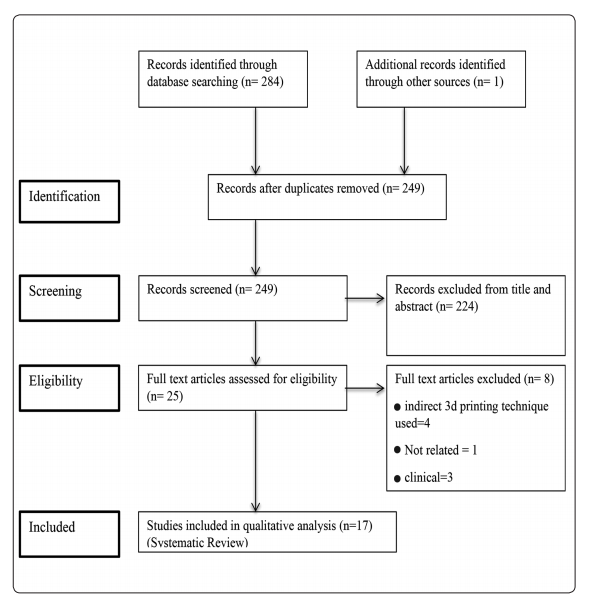
PRISMA flow chart of the systematic review
“Of the included 17 articles, 6 articles (35.3%) only assessed the marginal fit accuracy, one article (5.9%) only assessed the internal fit accuracy and 10 articles (58.84%) assessed both the marginal and internal fit accuracy,” explained Azer and Shahin. “Thirteen articles (76.5%) used single crown frameworks, 3 articles (17.6%) used fixed-partial-denture frameworks and only one article (5.9%) used both single crown and fixed partial-denture frameworks for the fit accuracy assessment.”
Cobalt – Chromium (Co-Cr) was used in all articles reviewed: a total of 14 studies employed direct metal laser sintering technique (DMLS), while 3 used selective laser sintering technique (SLS). Fabrication methods for comparing fit accuracy with laser sintering varied between:
- Lost wax method
- Wax pattern milling using CAD/CAM technology
- 3D printing of wax/resin pattern
“Among other techniques, milling of Co-Cr metal frameworks using CAD/CAM technology was used in 7 articles,” stated the authors. “Only one article used CAD/CAM zirconia milling.”
Methods used for both marginal and internal fit evaluation included:
- Silicone replica approach
- 3D replica approach
- Internal microscopic examination after cementation and sectioning of the specimen
- External microscopic examination of the marginal area
- Silicone impression weighing approach
- Direct-sight approach
Marginal and internal fit are the critical elements for success in fixed restoration, while just ‘marginal’ inaccuracies may cause:
- Gingival inflammation
- Gingival recession
- Secondary caries below crown margins
“According to American Dental Association (ADA) Specification No. 8, a gap width ranging between 25 to 40 μm has been suggested as a clinical goal,” explained the authors. “Sulaiman et al reported that 100 μm is an acceptable gap for clinical use. McLean and von Fraunhofer on the other hand have suggested that 120 μm should be the limit for clinical use. Moldovan et al reported that a gap of 200–300 μm is also acceptable. However, several researchers consider the value of 120 μm proposed by McLean and von Fraunhofer to be the most suitable limit for clinical use.”
Variations in the studies—even for the same system—were attributed to possible differences in fabrication technique, scanning, study designs—to include shape of casts, abutment teeth, and measurements. Such variations, however, rendered it impossible for the researchers to analyze the systems of rank them regarding accuracy.
“However, almost all the measurements were well within the clinically acceptable range suggested by McLean and von Fraunhofer. There was an agreement between the studies that the used systems have the ability to yield restorations with a clinically acceptable fit,” concluded the researchers.
“While further research is necessary to optimize the process parameters and clinical applications, the laser sintering procedure provides an efficient and rapid method for digitally designing and manufacturing complex metal structures for crowns and FPDs.”
3D printing in used in the dental industry today for projects such as making new crowns and bridges, new dental ecosystem materials, and scanning technology for dental arches. What do you think of this news? Let us know your thoughts! Join the discussion of this and other 3D printing topics at 3DPrintBoard.com.
[Source / Images: ‘Fit of Laser Sintered Metal Restorations: A Systematic Review’]
Subscribe to Our Email Newsletter
Stay up-to-date on all the latest news from the 3D printing industry and receive information and offers from third party vendors.
Print Services
Upload your 3D Models and get them printed quickly and efficiently.


You May Also Like
Stratasys Makes Navy Parts for Trident Warrior 25
The US Navy’s Trident Warrior 25 is a live fire manufacturing exercise hosted by FLEETWERX, an organization that wants to bring together companies and academia to drive Navy innovation, along...
3D Printing for Preppers: Ruggedized 3D Printers for Austere Environments
3D printers are ideal for making improvised, emergency repairs in austere environments. If you need something now, or a new design to improve something quickly, 3D printing is ideal. It’s...
Researchers Evaluate Comfort and Stability of 3D Printed Applicators for Oral Cancer Therapy
Oral cancer is on the rise around the world, and it’s especially bad in developing countries, such as Pakistan, Sri Lanka, and India, which don’t have the necessary medical infrastructure...
Marco Valenzuela of Additive Design Studio Makes an Innovative 3D Printed Pipette
Marco Valenzuela is a designer who specializes in crafting innovative and new 3D printed products. Originally coming from the gaming world, his Additive Design Studio now is focused on using Additive...