The UoT’s Faculty of Applied Science and Engineering reported that the compact instrument uses a roller to dispense bioink that is composed of mesenchymal stromal cells (MSCs) — stem cells that differentiate into specialized cell types depending on their environment– which promotes skin regeneration and reduces scarring.
The project is led by Richard Cheng, a doctoral candidate at the Institute of Biomaterials and Biomedical ![]() Engineering (IBBME) of the UoT, and is under the supervision of Axel Guenther, an associate professor at the Department of Mechanical & Industrial Engineering of the UoT, and in close collaboration with Marc Jeschke, director of the Ross Tilley Burn Centre, and his team at Sunnybrook Hospital.
Engineering (IBBME) of the UoT, and is under the supervision of Axel Guenther, an associate professor at the Department of Mechanical & Industrial Engineering of the UoT, and in close collaboration with Marc Jeschke, director of the Ross Tilley Burn Centre, and his team at Sunnybrook Hospital.
The first prototype of the skin printer was unveiled in 2018, and its creators believed it was the first device of its kind to form tissue in situ, depositing and setting in place in two minutes or less.
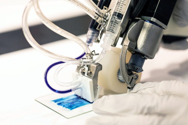
The handheld 3D skin printer developed by UoT Engineering researchers (Photo: Daria Perevezentsev)
As stated by the World Health Organization (WHO), burns are a global public health problem accounting for an estimated 180,000 deaths every year. In India alone, over 1,000,000 people are moderately or severely burnt every year; while in countries like Bangladesh, Colombia, Egypt, and Pakistan, almost 20 percent of children with burns have a temporary disability. Moreover, UoT reported that for patients with deep skin wounds, all three skin layers — the epidermis, dermis, and hypodermis — may be heavily damaged and the current preferred treatment is called split-thickness skin grafting, where healthy donor skin is grafted into the surface epidermis and part of the underlying dermis. However, they claim that split-thickness grafting on large wounds requires enough healthy donor skin to traverse all three layers, and sufficient graft skin is rarely available, leaving a portion of the wounded area ungrafted or uncovered, which results in poor healing outcomes.
Back in May 2018, Guenther indicated that “most current 3D bioprinters are bulky, work at low speeds, are expensive and are incompatible with clinical application.” The research team believed that their in-situ skin printer would be a platform technology to overcome the barriers for better patient solutions in burn injury while improving the skin-healing process.
“Previously, we proved that we could deposit cells onto a burn, but there wasn’t any proof that there were any wound-healing benefits — now we’ve demonstrated that,” indicated Guenther in a UoT statement.
Handheld 3D skin printer (Photo: University of Toronto)
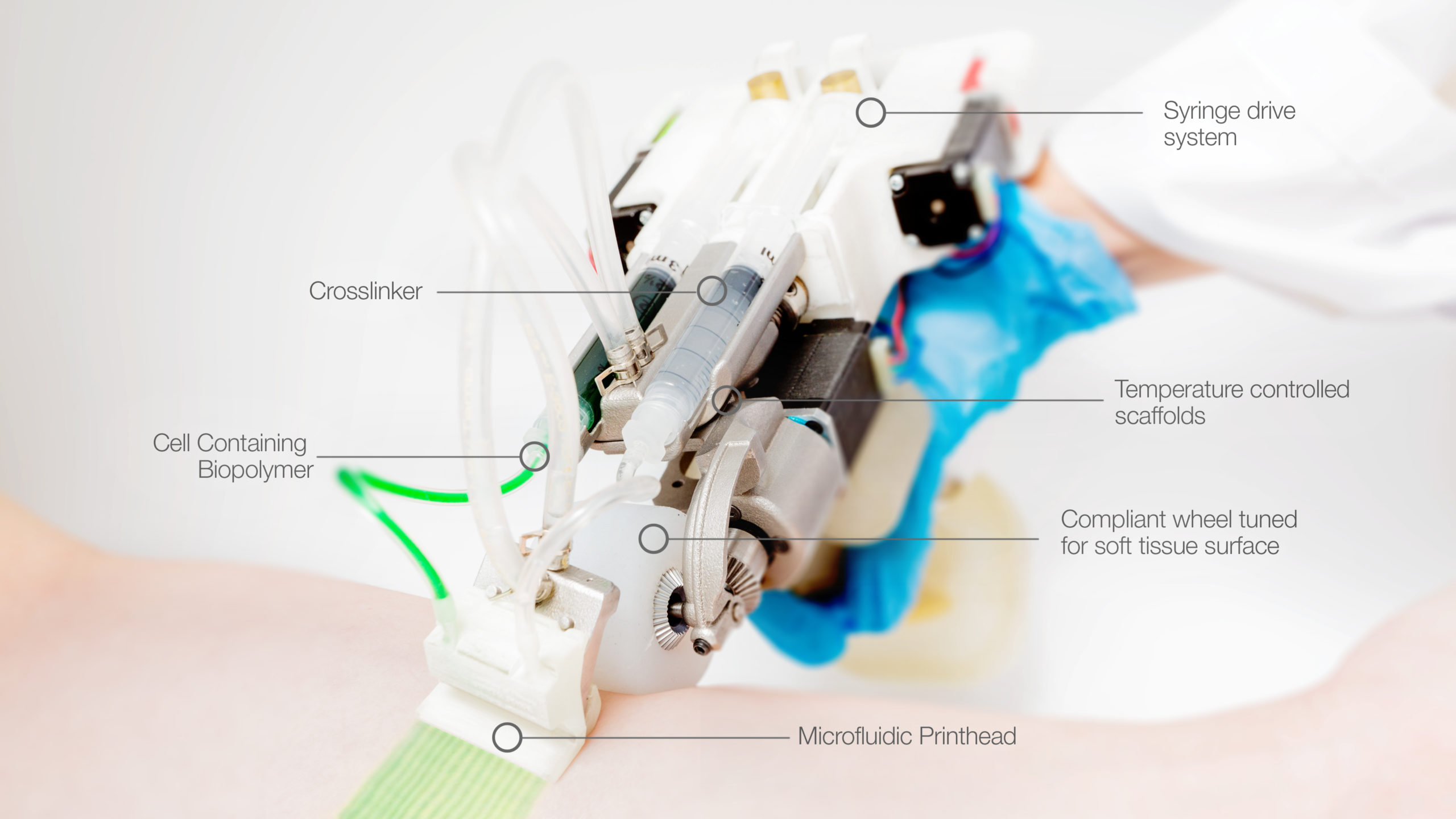
Since 2018, the printer has gone through 10 redesigns as the team moves towards a model they envision surgeons using in an operating room. The current prototype includes a single-use microfluidic printhead to ensure sterilization and a soft wheel that follows the track of the printhead, allowing for better control for wider wounds.
But how does it work? According to UoT, “two motors individually control the flow rate of the bioink and crosslinker filled syringes. The bioink then travels through the microfluidic cartridge and initiates gelation at the device exit after contact with the crosslinker. Rotational flexibility of the printhead allows homogenous printing on non-flat human surfaces, and a deformable wheel dissipates the pressure on the fragile wound bed while maximizing traction.”
The team chose enzymatically and thermally gelled fibrin and collagen biomaterials for their relevance in burn wound healing. Additionally, components in direct contact with either cells, biomaterials, or the wound, like the wheel, printer cartridges, and syringes, can be disposed of after a single use.
In porcine pre-clinical models of full-thickness burn, the team of researchers delivered “mesenchymal stem/stromal cell-containing fibrin sheets directly to the wound bed, improving re-epithelialization, dermal cell repopulation, and neovascularization, indicating that this device could be introduced in a clinical setting improving dermal and epidermal regeneration,” UoT states.
Victims of severe burn injury suffer from intense pain and require special treatment at specialized burn centers, usually involving medications, wound dressings, therapy, and surgery, to control pain, remove dead tissue, prevent infection, reduce scarring risk and regain function. The researchers at UoT’s one-step in situ formation of cell-containing biomaterial sheets using a handheld instrument that accommodates the topography of the wound is a breakthrough development that could help burn victim-survivors decrease a lot of the pain and suffering associated with post-burn treatment which leaves serious scarring, both physically and psychologically.