Treating Cancer Patients: Using 3D Scanning & Printing to Create the Bolus

In ‘A modern mold room: Meshing 3D surface scanning, digital design, and 3D printing with bolus fabrication,’ cancer researchers continue to seek better ways not just to find a cure, but to offer accurate, patient-specific treatment. Bolus walls are often used to protect the body as targeted areas are treated with radiotherapy. In this study, the researchers studied ten patients all requiring bolus in areas of the body with contours. To fight these challenges, the team designed a plan for 3D scanning, creating forms with PLA and then performing quality assurance tests for verification.
Historically, mold rooms have been used as dedicated space for patients receiving targeted treatments. And while 3D scanning has not been all that available for radiotherapy previously, that is changing now with greater affordability and accessibility to such technology—and especially FDM 3D printing. In this study, most patients were around 68 years old, with known tumors.
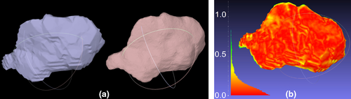
Post processing boluses created in Eclipse. a) A side by side comparison of an original bolus exported from the Eclipse TPS (left) and the smoothed bolus (right). Note that the step artifacts resulting from the 3 mm slice spacing are no longer apparent in the smoothed mesh. (b) For demonstration purposes, a detailed comparison between the two meshes is shown, illustrating that differences due to smoothing are negligible dosimetrically. The legend is in millimeters. The above analysis was not done routinely—a quicker method of verification was employed using a thickness measurement tool over the surface of the processed bolus. TPS, treatment planning system.
“The majority of patients (50%) had a diagnosis of basal cell carcinoma of the skin and two patients had a diagnosis of plasmacytoma,” stated the researchers. “Seven patients had superficial tumors involving the skin. Different dose and fractionation schedules were used depending on the pathology. Six patients were treated with megavoltage photons and the remaining four were treated with electrons.”
“An optical surface scan was acquired in three of seven superficial tumors for generating a 3D printed bolus; for the remaining patients CT scan data were used.”
For patients receiving CT scans for contour acquisition, the medical team used the Eclipse treatment planning system (TPS), and MeshMixer for more complex geometries. This meant that four out of ten pieces were created via Eclipse, and the rest with MeshMixer. Overall, the purpose was to ‘get the treatment geometry as close as possible to a water tank,’ affording the proper buildup.
During treatment, the medical professionals reported no ‘significant issues.’ Patients did not report discomfort using the 3D printed bolus, and therapists confirmed a good fit. It is also quite interesting to note that during the study no bolus pieces needed adjustment.
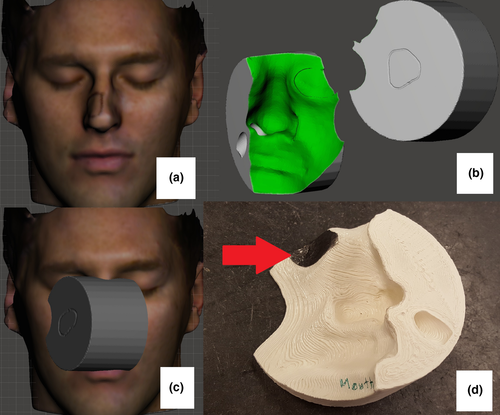
The design process for an electron bolus. a) Optical scan of volunteer for MeshMixer bolus design. b) Design, including flat surface on beam entrance side, breathing tunnel and eye shielding recess. c) Treatment geometry. d) printed bolus, with lead shielding in place (red arrow).
“This study demonstrates that 3D scanning, digital design, and 3D printing can be safely and effectively utilized to modernize bolus fabrication. The patients included here spanned a broad range of disease and treatment sites,” concluded the researchers.
“To ensure this new technology was implemented safely, QA tests were developed which are quick, quantitative, and thorough. Our process can be easily adopted by other centers since there is no reliance on equipment or software that is prohibitively costly. Both quantitative and qualitative data presented suggest that this design and fabrication process will enhance treatments requiring accessories in body regions with complex contours.”
3D printing is becoming more common not only in the medical field but in research for many diseases with the help of 3D printed models which may indicate tumors and more, but also in allowing for innovative drug delivery systems, and even 3D printed medications. What do you think of this news? Let us know your thoughts; join the discussion of this and other 3D printing topics at 3DPrintBoard.com.

Design process for a photon bolus. a) Knee lesion with affected skin marked with wire. b) Bolus designed in Eclipse before processing with MeshMixer. c) Printed bolus.
Subscribe to Our Email Newsletter
Stay up-to-date on all the latest news from the 3D printing industry and receive information and offers from third party vendors.
Print Services
Upload your 3D Models and get them printed quickly and efficiently.


You May Also Like
3D Printing Financials: 3D Systems Returns to Growth in Q1 2026
3D Systems (NYSE: DDD) reported one of its strongest quarters in recent years, showing signs that the company may finally be moving past the tough slowdown that has weighed on...
3D People Case Study Details Development of 3D Printed POV Camera Rig
A POV, or Point of View, camera rig, is a wearable support system that helps filmographers capture first-person footage, making the images more immersive. Some good examples of movies shot...
3D Printing News Briefs, May 2, 2026: Soft Robots, Agricultural Waste, & More
In this weekend’s 3D Printing News Briefs, we’ll start off with a multi-laser metal powder bed fusion 3D printer and post-processing news. We’ll end with research into soft robotics and...
Industrial Applications on Display at RAPID 2026: CERATIZIT & 3D Systems
Applications are where it’s at in the additive manufacturing (AM) industry. At the recent RAPID+TCT in Boston, I met with a few companies to learn about some of their very...