
3D Printing for Preoperative Management of Complex Orthopedic Cases


In the recently published, ‘Virtual preoperative planning and 3D printing are valuable for the management of complex orthopaedic trauma’ authors Abhishek Mishra, Tarun Verma, Abhishek Vaish, Riya Vaish, and Lalit Maini discuss the advantages of 3D printing medical models to prepare for orthopedic surgery.
Because orthopedic trauma can be so critical, and often fractures are so serious, planning must not only be strategic—but meticulous. When performed properly, preoperative training can cut down on time in the operating room as well as promising better outcomes statistically; however, as the authors discuss, preoperative training is not used as much as it should be—or even as much as it is recommended. Although some reasoning behind that may be the amount of time and money required for many types of preoperative tools, the opportunity for residents to learn and train is invaluable.
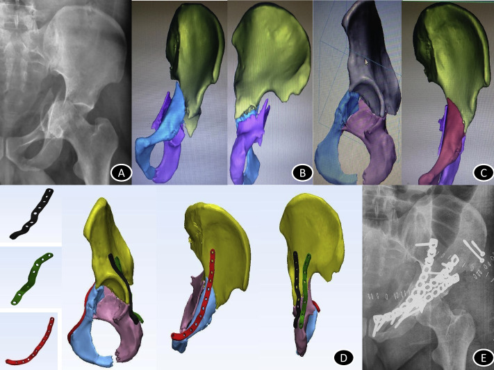
Surgical planning of a complex acetabular fracture case. (A) Preoperative X-ray; (B) 3D model of acetabular fracture; (C) Virtual reduction of fracture done; (D) Pre-contoured plate designed over reduced acetabular fracture; (E) Postoperative X-ray.

Virtual preoperative planning of a malunited tibial plateau fracture. (A) Preoperative X-ray of malunited tibial plateau fracture; (B) 3D reconstruction from non-contrast computed tomography; (C) Virtual 3D model designed from DICOM images; (D) Pre-contoured plate designed; (E) Reduction done virtually; (F) Postoperative X-ray.
For this study, the authors divided 91 complex orthopedic trauma cases into 12 different categories:
“(1) acetabular fracture, n=60; (2) nonunion distal tibia, n=1; (3) tibial plateau fracture, n=10; (4) cervical spine fracture, n=3; (5) combined convergent and divergent carpo-metacarpal fracture dislocation, n=1; (6) malunited fracture acetabulum, n=4; (7) shoulder fracture dislocation, n=4; (8) complex intra-articular elbow fracture, n=2; (9) posttraumatic elbow deformity, n=1; (10) comminuted distal radius fracture, n=2; (11) posttraumatic knee deformity, n=2 and (12) femoral head fracture, n=1.”
In all the cases, ultimately the surgeons were satisfied—both with intraoperative and postoperative outcomes. They found 3D printed models of the fractures to be helpful, and in the end upon filling out a questionnaire the main consensus was that the 3D printed training models rated a 4.5 out of 6.
While additive manufacturing is often used to fabricate bone models for bone regeneration cases, there are other materials commonly used such as ABS and PLA.
For young surgeons who need to train, both virtual planning and 3D printed models are an excellent opportunity to put both study and method to work. 3D images and models can be used to help in the creation of protheses too, often allowing for a much higher quality of life for patients.
“Jigs designed for placement of cervical pedicle screws save surgical time and reduce unnecessary exposure in image fluoroscopy. It is reported that each minute of exposure (60 shots) is equivalent to one computed radiography exposure or 4 Rads of radiation and that many periarticular fracture fixation scenarios require many minutes of these exposures,” concluded the researchers.
“Ultimately, VPP and 3DP reduce surgical duration and invasiveness and deliver better surgical outcomes. Although these also have an initial learning curve, with more experience of using these techniques, the time and efforts required become much lesser than initial cases. With improving technologies and advancements, the utility of these techniques is going to increase…”
While medical professionals in some areas of the world do have all the latest technology at their fingertip, many are just discovering the incredible benefits of 3D printing and medical models that can be used for so much more than just diagnosing a major health issue like a tumor. 3D printed medical models can also be used for treatment, training, preoperative training, and are often used in the operating today too. What do you think of this news? Let us know your thoughts! Join the discussion of this and other 3D printing topics at 3DPrintBoard.com.

Virtual preoperative planning of a comminuted proximal humeral fracture. (A) Preoperative X-ray; (B) Virtual 3D model designed from DICOM images; (C) 3D model printed with a 3D printer; (D) Pre-contoured plate template printed; (E) Pre-contoured plate designed; (F) Postoperative X-ray.
Subscribe to Our Email Newsletter
Stay up-to-date on all the latest news from the 3D printing industry and receive information and offers from third party vendors.
Print Services
Upload your 3D Models and get them printed quickly and efficiently.


You May Also Like
How One Artist Is Using 3D Printing to Tell Stories About the Ocean
Artist Kimberly Callas sees something different when she looks at a 3D printer. Where others see a machine for making parts, she sees a way to tell stories about the...
Bambu Lab Wants Home 3D Printing to Feel Less Like a Workshop with PLA Pure
As desktop 3D printers become increasingly common in homes, Bambu Lab is focusing attention on something beyond print speed and hardware features. This week, the company launched a new filament,...
AM Asia Watch: China Exported 2.46 Million 3D Printers in Four Months
China’s consumer 3D printer industry seems to be reaching a new level of global dominance. According to Chinese state media outlet China Global Television Network (CGTN), China exported 2.46 million...
Bambu Launches A2L: What the New Printer Reveals About Its Strategy
Bambu Lab continues its relentless march for 3D printing domination with the launch of the A2L. The 330 × 320 × 325 mm printer will have a nozzle temperature of...


