

Texas Researchers Find Success with 3D Printed Oral Stents

Texas researchers recently conducted a study regarding the use of 3D printed stents, outlining their findings in ‘Creating customized oral stents for head and neck radiotherapy using 3D scanning and printing.’ In this research, the authors compared three patients who received radiation for head and neck cancer with mouth opening tongue-depressing stents. They evaluated results as they were treated with models both from CT data and 3D scans converted into 3D printed models.
The oral radiation stent is in demand today over radiation therapy, helping patients to avoid serious issues like toxicity and radiation-induced oral mucositis (RIOM). The goal of the research study, ultimately, was to create a new workflow for manufacturing stents with 3D printing. Three patients, ages 35-66, two males and one female, were studied. Two of the patients had oropharyngeal cancer and one had paranasal sinus cancer. All had received radiation therapy previously and were sent to the Oral Oncology Department at The University of Texas MD Anderson Cancer Center (MDACC) to have the mouth opening tongue-depressing (MOTD) stents made—all under the approval of the Institutional Review Board at MDACC (protocol 2017–0269).
Articulated dental stone models from the dental laboratory were used, meant for production of conventional radiation stents. The researchers used these to scan maxillary and mandibular models on an EinScan Pro. According to the authors, these 3D scanned stents ‘successfully accommodated to patients’ teeth,’ while the CT models were not as accurate in replicating the bite.
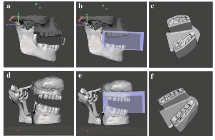
Workflow for digital stent design using CT-derived STL files (a–c), and the 3D-scanning method (d–f). a & d Mandibular rotation at Temporomandibular joint (TMJ) to achieve 20 mm inter-incisal opening; b & e Acquiring negative impression of the teeth; c & f Final completed stent.
Overall, the researchers stated that the 3D scans, CAD designs, and 3D printing made a great impact on efficiency in production. In comparison to CT imaging-based methods, the researchers were able to save substantially in time and effort, showing a ‘clear advantage’ in areas such as accuracy, reproducibility, and fit. There were several limitations, however, as obtaining the stone models took some effort, along with then having to rely on the models for evaluation instead of being able to fit the stents for the patients and get their feedback.
“Currently, we are conducting a clinical trial to comprehensively assess the efficiency of the proposed workflow in terms of time, labor, and patient-reported-outcomes (PROs). We also plan to apply our digital workflow to other stent designs such as the tongue lateralizing, mouth opening tongue-elevating, and lip protruding stents, which simply require swapping out digital components of the stent design,” concluded the researchers.
“Our results demonstrated the potential advantages of utilizing the 3D-scanning technology to overcome the inherent limitations associated with CT diagnostic imaging. The proposed workflow can be conveniently incorporated into radiation oncology practices, and future studies aiming to evaluate the clinical benefits of customized 3D-printed stents are warranted.”
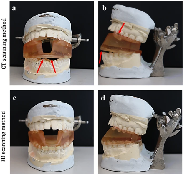
3D printed stent positioned on the dental stone model of a patient with an underbite. a & b Frontal and lateral view of the stent made from CT scan segmentation. Arrows point to areas where the stent does not fit into the model. c & d Frontal and lateral view of the stent made using the 3D scanning method.
3D printed medical models have made a huge impact in streamlining not just diagnosis and treatment of patients with serious conditions, but also in education for patients and their families, medical students, and surgeons who may be preparing for new or rarely performed procedures. What do you think of this news? Let us know your thoughts! Join the discussion of this and other 3D printing topics at 3DPrintBoard.com.
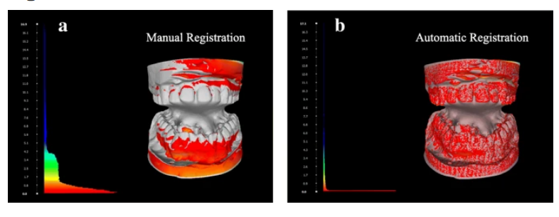
Hausdorff distance computation between the registered meshes and the base mesh, followed by color mapping by vertex quality. a manual registration b automatic registration using the ICP algorithm.
Subscribe to Our Email Newsletter
Stay up-to-date on all the latest news from the 3D printing industry and receive information and offers from third party vendors.
Print Services
Upload your 3D Models and get them printed quickly and efficiently.


You May Also Like
Bambu Launches A2L: What the New Printer Reveals About Its Strategy
Bambu Lab continues its relentless march for 3D printing domination with the launch of the A2L. The 330 × 320 × 325 mm printer will have a nozzle temperature of...
3D Printing News Briefs, May 30, 2026: RIMPAC 2026, Acquisition, Ceramic Implants, & More
We’re kicking things off with materials news in this weekend’s 3D Printing News Briefs. Then it’s on to a hybrid manufacturing system for a maritime exercise, an expansion of industrial...
3D Printing News Briefs, May 28, 2026: Continuous Fiber Reinforcement, Bioprinted Trachea, & More
In today’s 3D Printing News Briefs, America Makes announced the winners of its JAQS-SQ Project Call. Axtra3D is partnering with Keystone Industries to expand its dental material ecosystem, while BigRep...
Peopoly Unveils $15,000 Giga 800 FGF 3D Printer for Large Pellet Prints
Peopoly is known for its Magneto X, a printer that eschews belts and uses linear motors instead. The company also has a vat polymerization system, and now is moving into FGF...


