FluidForm & Carnegie Mellon: Closer Than Ever to Bioprinting a Human Heart

While the realm of bioprinting has continued to expand significantly in the past few years, allowing for better sustainability of cells in the lab—and the bioink used to print them—the end goal is almost always to get just that much closer to 3D printing organs, meaning the potential for enormous change in the current medical culture of patients suffering and often dying while waiting for transplants. Now, with a collaboration between startup FluidForm and Carnegie Mellon University, scientists may actually be much closer than ever to bioprinting a human heart.
Findings on the subject were published recently in the August 2nd edition of Science regarding work by the Carnegie Mellon University research team, made up of co-first authors and FluidForm co-founders Andrew Lee and Andrew Hudson, and the nine members of the Carnegie Mellon team. They have just developed an advanced version of Freeform Reversible Embedding of Suspended Hydrogels (FRESH) technology which allows them to 3D print collagen. With these new advancements, the scientists can fabricate cardiac components, including tiny blood vessels, valves, and beating ventricles.
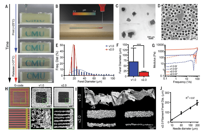
(A) Time-lapse sequence of 3D bioprinting of the letters “CMU” using FRESH v2.0. (B) Schematic of acidified collagen solution extruded into the FRESH support bath buffered to pH 7.4, where rapid neutralization causes gelation and formation of a collagen filament. (C and D) Representative images of the gelatin microparticles in the support bath for (C) FRESH v1.0 and (D) v2.0, showing the decrease in size and polydispersity. (E) Histogram of Feret diameter distribution for gelatin microparticles in FRESH v1.0 (blue) and v2.0 (red). (F) Mean Feret diameter of gelatin microparticles for FRESH v1.0 and v2.0 [N > 1200, data are means ± SD, ****P < 0.0001 (Student t test)]. (G) Storage (Gʹ) and loss (Gʺ) moduli for FRESH v1.0 and v2.0 support baths showing yield stress fluid behavior. (H) A “window-frame” print construct with single filaments across the middle, comparing G-code (left), FRESH v1.0 (center), and FRESH v2.0 (right). (I) Single filaments of collagen showing the variability of the smallest diameter (~250 μm) that can be printed using FRESH v1.0 (top) compared to relatively smooth filaments 20 to 200 μm in diameter using FRESH v2.0 (bottom). (J) Collagen filament Feret diameter as a function of extrusion needle internal diameter for FRESH v2.0, showing a linear relationship.
“We now have the ability to build constructs that recapitulate key structural, mechanical, and biological properties of native tissues,” said Prof. Adam Feinberg, CTO and co-founder, FluidForm, and Principal Investigator, Regenerative Biomaterials and Therapeutics Group, Carnegie Mellon, where the research was done. “There are still many challenges to overcome to get us to bioengineered 3D organs, but this research represents a major step forward.”
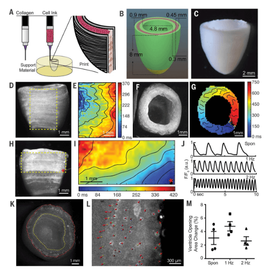
Created from MRI data, the FRESH bioprinted hearts were comprised of small cardiac ventricles printed with human cardiomyocytes that demonstrated the following:
- Synchronized contractions
- Directional action potential propagation
- Wall-thickening up to 14 percent during peak systole
“We found that FRESH 3D-bioprinted hearts accurately reproduce patient-specific anatomical structure as determined by micro–computed tomography. Cardiac ventricles printed with human cardiomyocytes showed synchronized contractions, directional action potential propagation, and wall thickening up to 14% during peak systole,” state the researchers in their paper, “3D bioprinting of collagen to rebuild components of the human heart.”
Other obstacles are still present though, as scientists wrestle with generating the billions of cells needed for 3D printing larger tissues.
“Although the 3D bioprinting of a fully functional organ is yet to be achieved, we now have the ability to build constructs that start to recapitulate the structural, mechanical, and biological properties of native tissues,” stated the researchers in their paper.
While previously scientists have projected bioprinting of organs in the very near future, it has proven to be much more difficult than anticipated, obviously. Milestones continue to be surpassed, but challenges continue to erupt—mainly in keeping cells alive in coordination with the proper technology—meaning materials, software, and hardware continue to be a significant focus. With FRESH, a temporary support gel is used to 3D print collagen scaffolds, driven by a rapid pH change that drives collagen self-assembly. There is promise not only for 3D printing hearts, however, but a range of different tissues and organs in the future.
“FluidForm is extraordinarily proud of the research done in the Feinberg lab” said Mike Graffeo, CEO, FluidForm. “The FRESH technique developed at Carnegie Mellon University enables bioprinting researchers to achieve unprecedented structure, resolution, and fidelity, which will enable a quantum leap forward in the field. We are very excited to be making this technology available to researchers everywhere.”
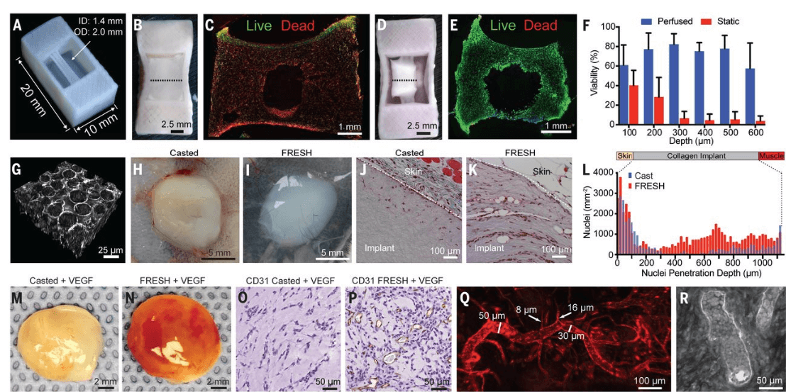
(A) FRESH-printed collagen tube construct. (B) C2C12 cell and collagen gel mixture cast around the collagen tube and static-cultured for 5 days. (C) Cross section of the tissue from (B) stained for live (green) and dead (red) cells. (D) C2C12 cell and collagen gel mixture cast around the collagen tube and perfused for 5 days. (E) Cross section of the tissue from (D) stained for live and dead cells. (F) Percent cell viability as a function of depth from the top surface of the tissues from the static and perfused collagen tube constructs [N = 3, data are means ± SD, *P < 0.05 (two-way ANOVA followed by Bonferroni multiple-comparisons posttest)]. (G) Multiphoton imaging showing microscale porosity in FRESH-printed collagen constructs after removal of the gelatin microparticle support bath. (H and I) Collagen constructs cast (H) and FRESH-printed (I) without VEGF 7 days after subcutaneous implantation. (J and K) Masson’s trichrome staining to visualize cells (red) and collagen (blue) in cast (J) and FRESH-printed (K) collagen disks after 7 days of subcutaneous implantation. (L) Cell density after implantation as a function of depth for the cast and FRESH-printed collagen disks. (M and N) Collagen constructs cast (M) and FRESH-printed (N) with VEGF (100 ng/ml) 10 days after subcutaneous implantation. (O and P) CD31 staining (brown) and cells (blue) of cast (O) and FRESH-printed (P) collagen disks doped with VEGF (100 ng/ml) after 10 days of subcutaneous implantation. (Q) Host vascular infiltration of vessels (diameter 8 to 50 μm) in the FRESH-printed collagen disk labeled by lectin tail vein injection (red). (R) Multiphoton image 70 μm into the FRESH-printed construct, showing red blood cells within the lumen of the microvasculature.
(A) FRESH-printed collagen tube construct. (B) C2C12 cell and collagen gel mixture cast around the collagen tube and static-cultured for 5 days. (C) Cross section of the tissue from (B) stained for live (green) and dead (red) cells. (D) C2C12 cell and collagen gel mixture cast around the collagen tube and perfused for 5 days. (E) Cross section of the tissue from (D) stained for live and dead cells. (F) Percent cell viability as a function of depth from the top surface of the tissues from the static and perfused collagen tube constructs [N = 3, data are means ± SD, *P < 0.05 (two-way ANOVA followed by Bonferroni multiple-comparisons posttest)]. (G) Multiphoton imaging showing microscale porosity in FRESH-printed collagen constructs after removal of the gelatin microparticle support bath. (H and I) Collagen constructs cast (H) and FRESH-printed (I) without VEGF 7 days after subcutaneous implantation. (J and K) Masson’s trichrome staining to visualize cells (red) and collagen (blue) in cast (J) and FRESH-printed (K) collagen disks after 7 days of subcutaneous implantation. (L) Cell density after implantation as a function of depth for the cast and FRESH-printed collagen disks. (M and N) Collagen constructs cast (M) and FRESH-printed (N) with VEGF (100 ng/ml) 10 days after subcutaneous implantation. (O and P) CD31 staining (brown) and cells (blue) of cast (O) and FRESH-printed (P) collagen disks doped with VEGF (100 ng/ml) after 10 days of subcutaneous implantation. (Q) Host vascular infiltration of vessels (diameter 8 to 50 μm) in the FRESH-printed collagen disk labeled by lectin tail vein injection (red). (R) Multiphoton image 70 μm into the FRESH-printed construct, showing red blood cells within the lumen of the microvasculature.
Subscribe to Our Email Newsletter
Stay up-to-date on all the latest news from the 3D printing industry and receive information and offers from third party vendors.
Print Services
Upload your 3D Models and get them printed quickly and efficiently.


You May Also Like
Where the Money Is Going: The New Infrastructure Landscape
This is Part 1 of a two-part PRO series examining where infrastructure investment is flowing and how those trends are reshaping manufacturing, energy, logistics, and additive manufacturing. Part 2, by...
3D Printing & Drone Dominance: Speed, Performance, and Derisking the Supply Chain
A shift is underway in drone manufacturing. Government programs like the U.S. Department of War’s Drone Dominance, a $1.1 billion effort to deliver low-cost, one-way attack (OWA) sUAS at scale,...
3D Printing Financials: Stratasys Bets on Defense and Drones as Printer Sales Slow
Stratasys (Nasdaq: SSYS) started 2026 with lower revenue and a larger loss as customers continued to slow down spending on new 3D printers. Still, the company pointed to stable recurring...
INDOPACOM Advanced Manufacturing Team Saves Thousands Per Week at Joint Exercise in the Philippines
In the summer of 2025, the US Indo-Pacific Command (INDOPACOM) opened a new advanced manufacturing facility at Schofield Barracks, the Hawaiian home of the US Army’s 25th Infantry Division. INDOPACOM...