

Researchers Rely on 3D Printed Models & Surgical Guides for Pediatric Orthopedic Surgery


Medical researchers and orthopedic surgeons in Taiwan at Kaohsiung Veterans General Hospital continue to explore better ways to heal bones and manage defects, with their findings outlined in the recently published ‘Anatomic three-dimensional model-assisted surgical planning for treatment of pediatric hip dislocation due to osteomyelitis.’
While bone defects are already a challenge to manage, obviously the problem is compounded in children, with smaller bones being even more difficult to repair in surgery. Currently, there are few options for a good device meant for small bone repair during pediatric osteotomies—making it difficult for surgeons around the world to correct both subluxated hip joints and deformed femurs in children.
The authors (and surgeons) performed corrective surgery on a four-year-old boy with a post-osteomyelitis deformity. In preparing for the surgery, they relied on a 3D printed model of the bone for studying the condition, surgery and preparing the site for the appropriate implant. Because this type of surgery requires ‘meticulous planning,’ the doctors required both 2D and 3D assistance, in the respective forms of axial images and 3D virtual models of patient anatomies.
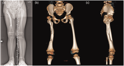
Radiographs taken before corrective surgery. (a) Triple film showing the proximal femur deformity with osseous recovery. Three-dimensional computed tomography image: (b) anteroposterior and (c) lateral views
As the surgeons examined the patient and reviewed the CT, they noticed a genu valgus deformity (more commonly known as a ‘knock-knee’ condition). Another corrective surgery was scheduled, with 3D CT imaging examined for bone tissue analysis. The surgeons realized, however, that the procedure would be more successful overall with a life-size 3D model. They were able to outline a patient-specific plan, also bringing in additional assistance from an orthopedic consulting firm focused around 3D orthopedics and ‘patient-specific instrumentation.’

Customized-to-patient three-dimensionally–printed guide. (a) The patient-specific guide for our patient. (b) Two resecting osteotomies can achieve optimal joint congruency and varus angle correction. (c) Correcting the femoral rotation would result in joint translation in both the coronal and axial planes
What was also very valuable to the surgery—and the outcome for the little boy involved—was that the surgeons could use the model to practice on, exercising ‘simulations of possible osteotomy options.’
“After a few osteotomy options had been analyzed, one osteotomy cut was made vertically to the femoral shaft on the subtrochanteric area, and another was made on the middle third of the femur to correct the bowing deformity of the midshaft,” stated the researchers. “Correction of femoral rotation can result in either joint translation in the coronal and axial planes or difficulty with fixation, both of which could be prevented with the help of the 3D model in the present case.”
The results of the surgeries were successful, with the patient able to stretch and begin other mobilization activity after four months.
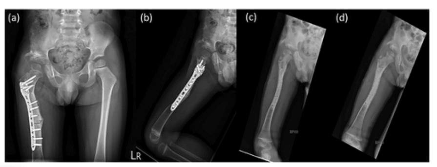
Postoperative (a) anteroposterior and (b) lateral views. Fifteen-month postoperative (c) anteroposterior and (d) lateral views
“The result of our case suggests that the use of 3D printing models improves the postoperative performance as shown by both physical function and radiological evidence,” stated the authors in the concluding discussion.
“The use of a 3D-printed patient-specific guide is a safe, modern, affordable, and promising method that offers advantages including a shorter surgical time, optimally positioned implant placement, acceptable alignment, and a probable lower rate of complications. The utilization of 3D-printed models for skeletal deformity surgery, especially complex and difficult pediatric surgery, provides superior precision and foreseeably better outcomes. We strongly believe that with the promotion of 3D printing methodology, models for preoperative planning may soon become the gold standard for pediatric deformity correction surgery.”
3D printing continues to make impacts in the area of healing bones, regeneration and planning for complex surgeries with a range of medical devices and models. What do you think of this news? Let us know your thoughts! Join the discussion of this and other 3D printing topics at 3DPrintBoard.com.
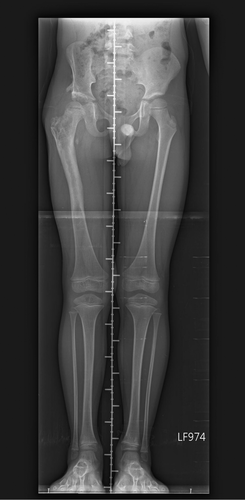
Triple film at 2-year postoperative follow-up showing no significant leg length discrepancy (<0.5 cm)
Subscribe to Our Email Newsletter
Stay up-to-date on all the latest news from the 3D printing industry and receive information and offers from third party vendors.
Print Services
Upload your 3D Models and get them printed quickly and efficiently.


You May Also Like
The Seminal Moment: Creality’s IPO Analysis & Possible Effects
Something super important happened just a few days ago, and too few people paid attention. Creality, a pioneer in low-cost desktop material extrusion printers, went public. Creality is now listed...
As Longevity Gains Momentum, Rem3dy Health Raises £14 Million for 3D Printed Nutrition
Longevity hack or healthcare trend? The answer may depend on who you ask, but investor interest in personalized nutrition is growing as consumers search for the next longevity hack. Now,...
3Dnatives to Present ADDITIV Metals 2026: Resolving Key Barriers to Scaling Metal Additive Manufacturing
As the metal additive manufacturing sector prepares for a massive leap—with market valuations expected to climb from $6.02 billion to $7.02 billion this year—the industry is shifting its focus from...
Stratasys Dental’s Negar Movahed Says They’re “Open for Partnerships”
According to “3D Printing for Dentistry 2025: Market Study and Forecast” by AM Research, the dental 3D printing market generated $5.2 billion in revenue in 2024—that’s nearly one third of...


