3D Printed Scaffolds Evaluated for Mandibular Reconstruction


In ‘Digital Design, Analysis and 3D Printing of Prosthesis Scaffolds for Mandibular Reconstruction,’ we learn more about strides in the medical field regarding mandibular surgeries, reconstruction, the potential for current and future improvements in implants, as well as understanding more about the obstacles today and how they can be overcome.
 3D printing is invaluable to the medical field overall due to the ability to customize nearly everything—and offer patient-specific care to individuals who may have faltered under previous one-size-fits-all restrictions. Customized implants especially are offering new options to patients, as porous, lightweight structures can be created to aid in reconstruction and the eventual return to normal functions such as chewing and swallowing nutrients. For this study, Saudi researchers from King Saud University created two different customized scaffold designs (a top and bottom plate, and an inner porous plate) which they later evaluated regarding both usefulness and aesthetic appearance.
The authors describe the perfect scaffold as one that is highly porous, without cracks, and fully biocompatible. Previous studies have shown that titanium is highly effective as a material but may also lead to deterioration of mechanical properties and eventual resorption or failure of the implant. 3D printing has offered potential in reconstruction, but also rehabilitation, and surgery too.
“Among several 3D printing techniques, electron beam melting (EBM) has been regarded as the fast and successful method for the fabrication of titanium medical implants from computer-aided design (CAD) models with Food and Drug Administration (FDA) and Conformité Européene (CE) approval,” stated the researchers.
Until now, the researchers state that there has been no clear research regarding mandibular reconstruction or biomechanical properties, stability and integrity, or testing of related scaffolds and structures. Here, they studied a patient, at 40 years old, suffering from deformities and a ‘lesion in the left mandibular area.’ The patient was treated in an emergency department initially, beginning with a series of CT scans uncovering a mandibular continuity defect, and substantial loss of bone.
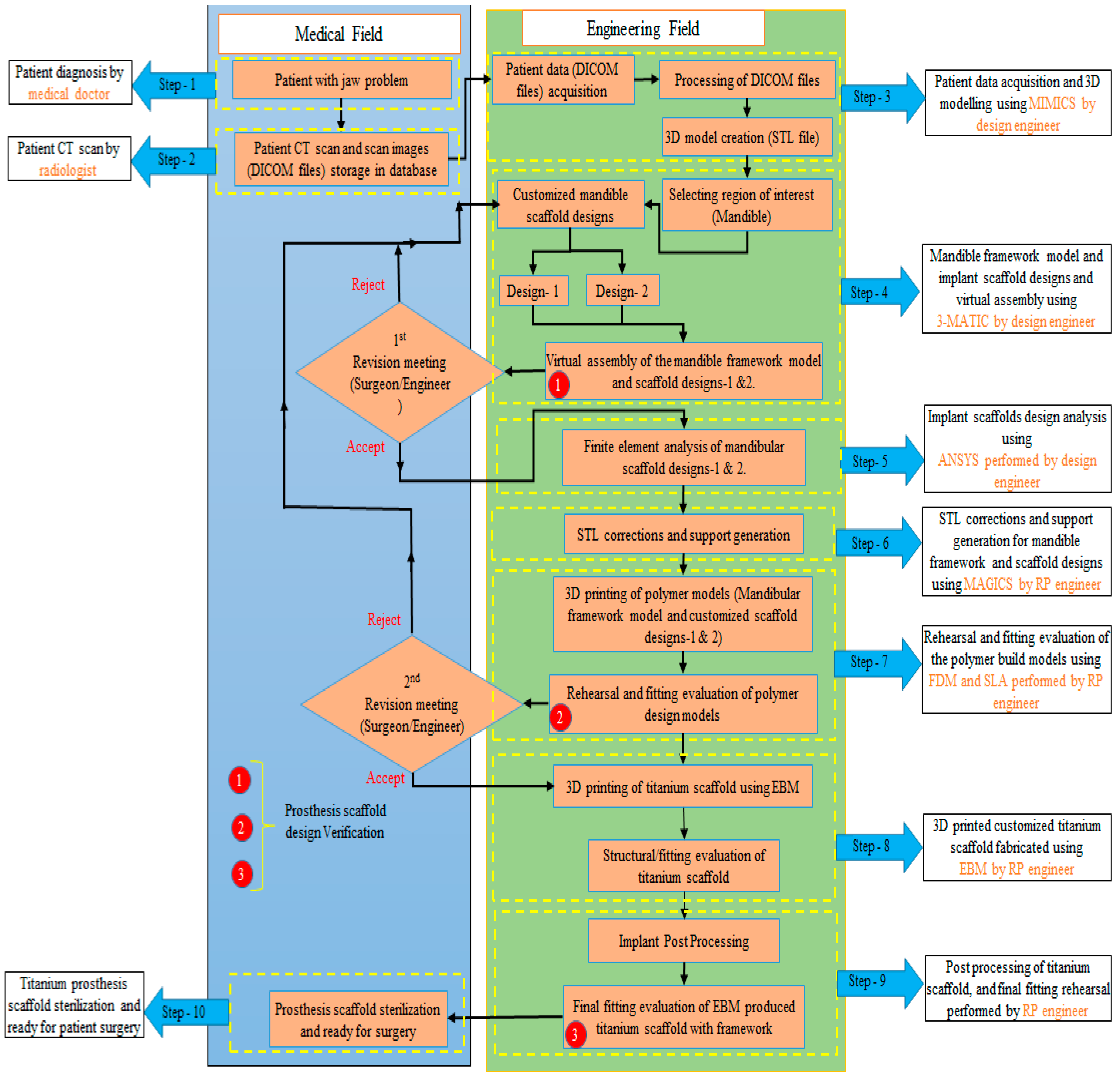
The proposed methodology for design, analysis and fabrication of customized mandibular prosthesis scaffolds. Note: The red circles indicate the formal meetings between the engineering and medical department for scaffold design verification and evaluation.
The images were converted into 3D files using Materialise Mimics software, and then an FDM 3D printer was used with ABS material to create the framework model. A Formlabs 2 SLA 3D printer was then used for fabrication of the mandibular prosthesis scaffold. The researchers evaluated the structures for weight, integrity, and accuracy, using a micro-CT scan.
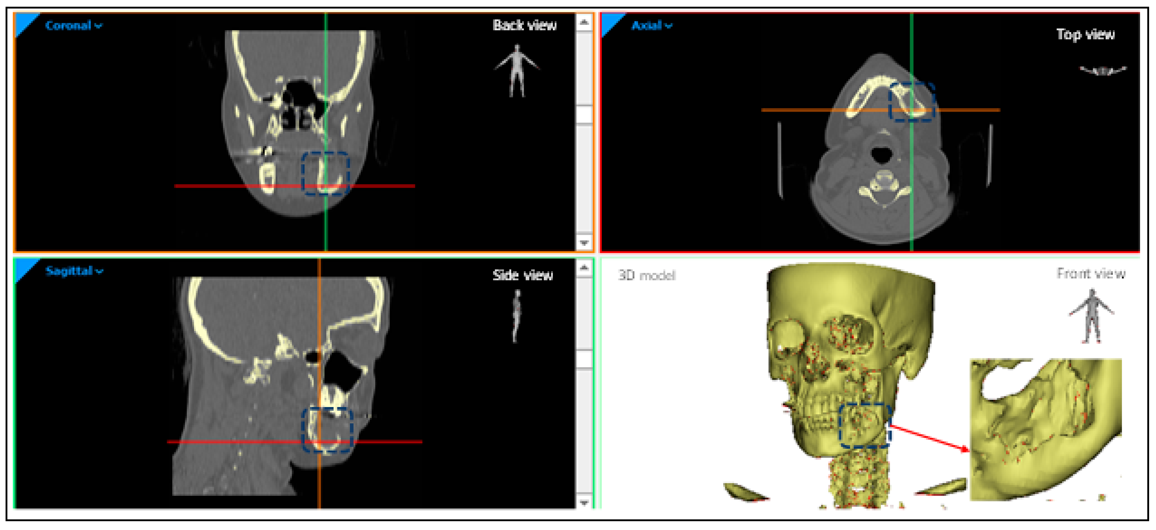
Patient anatomical model depicting the tumor region in different planes.
“Eventually, depending on the FEA, weight analysis and fitting accuracy evaluation, it can be inferred that the scaffold with the top and bottom porous plate is more favorable for bone reconstruction as compared to scaffold with the inner porous implant and can successfully be employed in the reconstruction of the defective mandible. Indeed, it can be asserted that the employment of prosthesis scaffolds in mandibular reconstruction satisfies the sustained need of lighter implants with accurate fitting and lesser surgical time and minimal revisions,” concluded the researchers.
“It is mandatory that the research in this area should continue in the future for acquiring further innovative implant designs and reconstruction methods. The authors would like to expand this work by introducing new designs with different porous structures and analyzing them for their strength and accuracy in mandible restoration. In addition, the authors would like to extend this work by including an extensive clinical (in-vivo) study in the future.”
Mandibular reconstruction can be a challenging endeavor, but when successful it offers incredible value to patients who may be having trouble chewing, as well as enduring understandable self-consciousness due to a defect or tumor removal.
This is big topic of research and has also led to numerous innovations via 3D printing, from alveolar ridge augmentation to mandibular grafts, and implants for cancer patients. What do you think of this news? Let us know your thoughts! Join the discussion of this and other 3D printing topics at 3DPrintBoard.com.
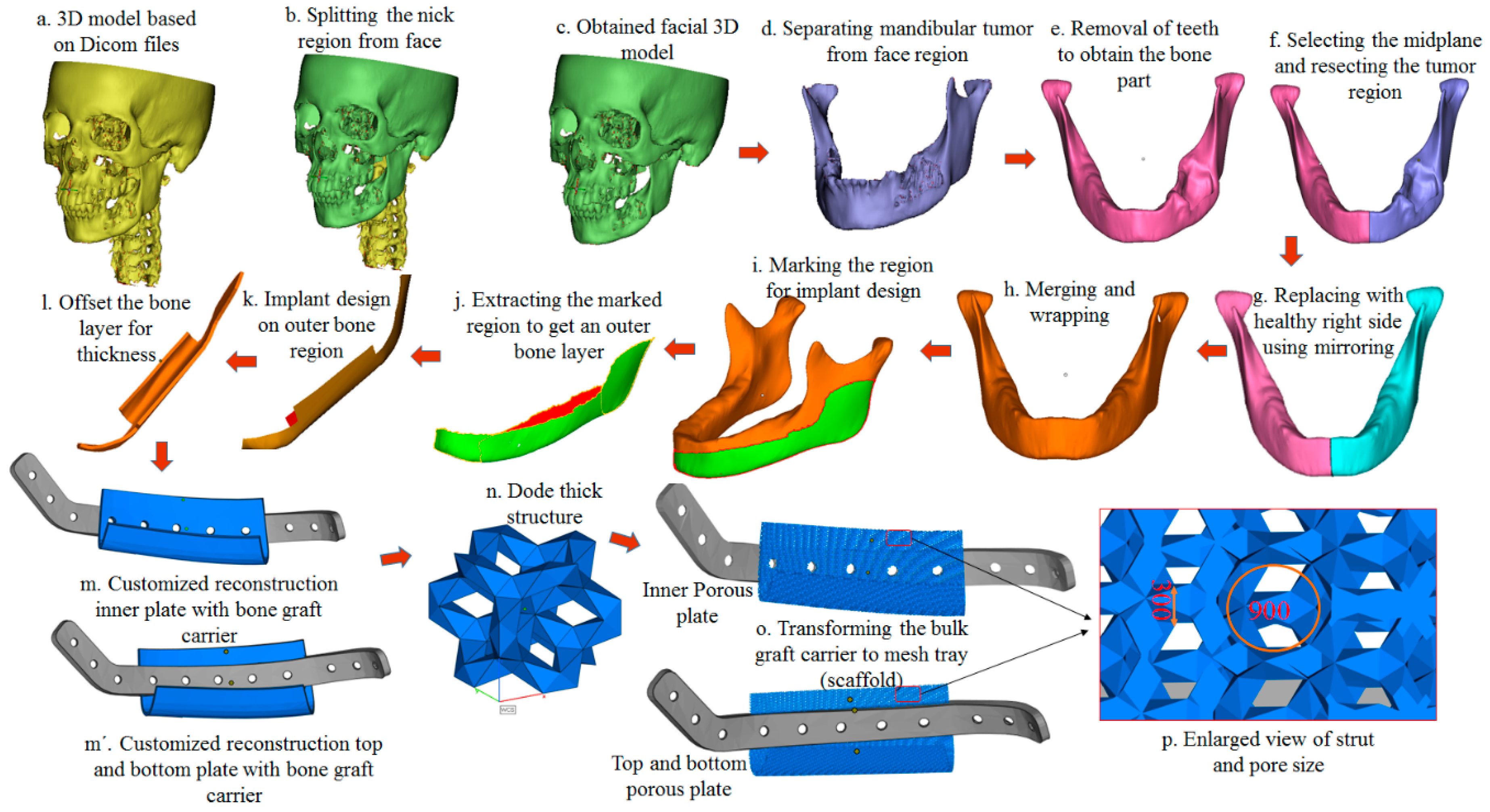
Sequence of steps in the design of customized prosthesis scaffold (implant) for mandibular defects.
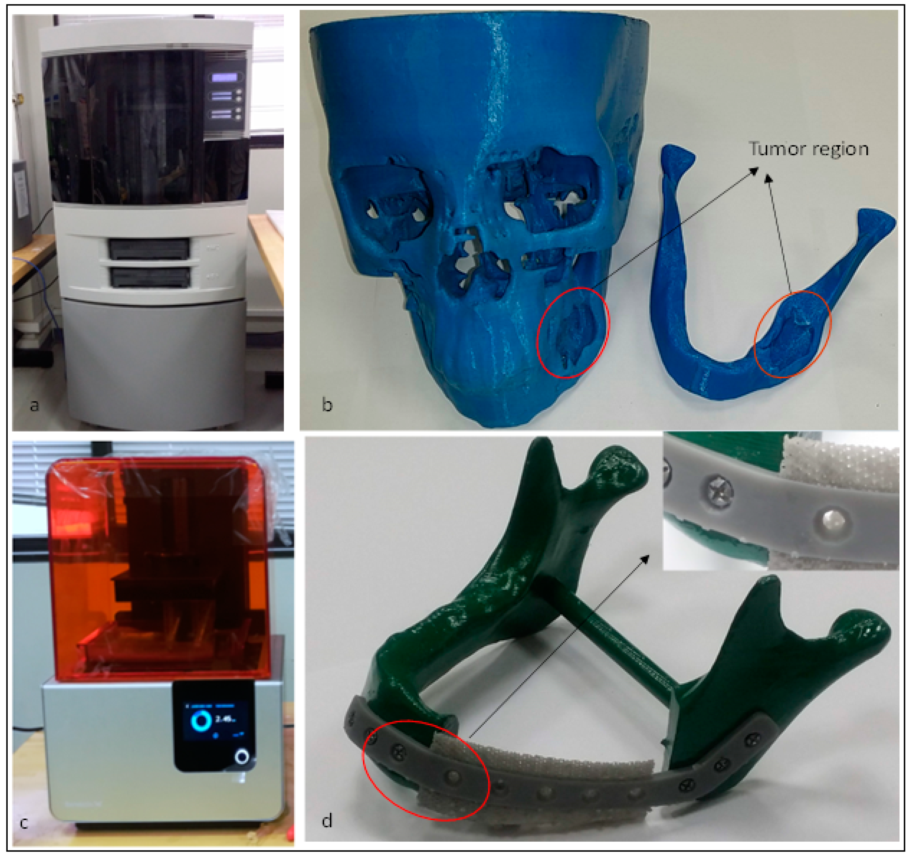
(a) Fused Deposition Modeling machine with its fabricated polymer model (b) indicating the tumor region and (c) SLA machine and its produced mandibular scaffold (d) with a close-up view.
Subscribe to Our Email Newsletter
Stay up-to-date on all the latest news from the 3D printing industry and receive information and offers from third party vendors.
Print Services
Upload your 3D Models and get them printed quickly and efficiently.


You May Also Like
3D Printing in Drones Could Reach $900 Million by 2034, AM Research Report Says
For years, additive manufacturing has searched for applications where its advantages clearly outweigh the limits of traditional production methods. Now, according to a new report from Additive Manufacturing Research (AM...
Largest Publicly Announced, Single Order in EOS History: Beehive Industries Spends $50M on M4 ONYX 3D Printers
Earlier this year, Beehive Industries received a $29.7 million contract to produce its Frenzy 6 and Frenzy 8 engines for the US Air Force. The metal additive manufacturing (AM) user...
3D Printing News Briefs, June 10, 2026: Grand Opening, Photoresins, Footwear, & More
We’re starting with some exciting news in today’s 3D Printing News Briefs: Stratasys just celebrated the opening of its new North American headquarters in Minnesota. Moving on, Nanoscribe is scaling...
Formlabs Launches the X1: Let’s Pack It In?
I’d like to share my disappointment. When Formlabs teased that something big was coming, I really hoped the firm would finally get around to making CNC machines, as it always...