Researchers Create Patient-Specific 3D Printed Meniscus Prototype

 If you can sprint off for a pleasant morning run without wincing or heading for the pain reliever afterward, consider yourself lucky—and the envy of many who suffer from current or healing meniscus strain and injuries. Researchers at the Istituto Ortopedico in Rizzoli, Bologna, recently published findings in ‘Patient-specific meniscus prototype based on 3D bioprinting of human cell-laden scaffold,’ attempting to improve on current methods for making tissue repairs and replacement.
Authors G. Filardo, M. Petretta, C. Cavallo, L. Roseti, S. Durante, U. Albisinni, and B. Grigolo used real MRI scans from one patient and converted to the data into an .stl file, then proceeding to create a model from which to make the meniscus prototype and resulting scaffolds.
In operating on patients suffering from classic meniscus tears, the surgeon’s goal is usually to preserve as much healthy tissue as possible, while some may require total meniscectomies. Transplants today, however, are rife with all the typical challenges such as problems with rejection of the tissue, mismatch, and other issues such as impaired cellular infiltration. In this project, the researchers examined other challenges with scaffolds that have been previously created too, stating that only two types of artificial meniscus have made it to the clinical arena—one of which is a collagen implant, and the other constructed out of polyurethane and polycaprolactone. While they were both evaluated to be safe, and offered good results, the researchers point out that the ‘mismatch’ between implants and ‘patient-specific lesion areas’ is still a central problem.
“… it has been demonstrated that even small changes in implant positioning may affect contact pressure and joint stress,” stated the research. “To address the limits of meniscus implantation, and to optimize the restoration of meniscal function and joint integrity over time, implants could be developed with an enhanced biological potential and patient-specific sizing to meet individuals’ joint requirements.”
The researchers harvested bone marrow from a patient already scheduled for autologous cell transplantation, and watched cells expand and continue to thrive even after a week. The researchers used the 3D printed model of the knee to assist in reconstruction of the meniscus. They used a series of 2D cross sections to create tool paths, using LifeInk 200 bio-ink as the material for printing cells.
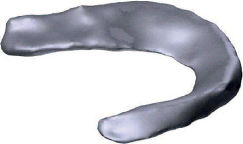
.stl model of a human meniscus.
“The selected bio-ink presented good printability and shape fidelity, allowing the fabricated tissue, obtained by means of a microvalve-based inkjet dispensing technique, to mimic the anatomical model morphology,” stated the researchers. “This ‘cell-friendly’ technology allowed MSCs included in the bio-ink to be homogeneously distributed within the construct.”

Photograph of a custom-made, human, cell-laden, high-density collagen type I meniscus prototype after mesenchymal stem cells were embedded. The printing process was performed at room temperature in a Petri dish filled with culture medium and kept at 37°C.
After five days, only half of the cells were still alive, creating concerns about cell viability. Surprisingly though, almost the entire second half of the cells still alive were viable after 28 days.
“… these cells were able to grow and to colonize the biomaterial, demonstrating that the bioprinted collagen-based hydrogel scaffold provides a good microenvironment for the viability and proliferation of MSCs,” stated the researcher.
Overall, the research showed that this type of bioprinting shows the potential for tissue engineering with ‘anatomical precision.’ They do note, however, that several issues must be scrutinized in the future: density, cell encapsulation inside collagen gels, and collagen gel pore size.
“The prototype described in this study showed the biological potential of 3D bioprinting technology in providing an anatomically shaped, patient-specific construct with viable cells on a biocompatible material,” concluded the authors. “This study could act as the starting point for future developments of this custom-made, collagen-based, tissue-engineered structure, which could aid the optimization of implants designed to replace damaged menisci.”
Cartilage is a hot commodity for humans, and especially as the wear and tear of age begins to show, with this happening earlier for some of us. 3D printing shows huge potential to help with repairs, however, with new and promising methods for repairing defects, curing arthritis, bioprinting with knee stem cells to make new cartilage, and more. Find out more about advances in meniscus repairs here. What do you think of this news? Let us know your thoughts; join the discussion of this and other 3D printing topics at 3DPrintBoard.com.
[Source / Images: Patient-specific meniscus prototype based on 3D bioprinting of human cell-laden scaffold]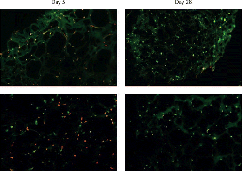
LIVE/DEAD images of cell-laden collagen type I gel scaffolds. Viable cells are in green and dead cells in red. The top row shows slides from total meniscus structure, while the bottom row shows slides from cubical constructs. Images are representative.
Subscribe to Our Email Newsletter
Stay up-to-date on all the latest news from the 3D printing industry and receive information and offers from third party vendors.
Print Services
Upload your 3D Models and get them printed quickly and efficiently.


You May Also Like
Scientists Create Stretchy 3D Printed Implants for High Blood Pressure Treatment
Researchers at Pennsylvania State University (Penn State) say they may have found a softer, less invasive way to treat severe high blood pressure. In a new study published in the...
Harvard’s Jennifer Lewis Lab Is 3D Printing Artificial Muscles That Twist and Bend on Demand
Researchers at Harvard John A. Paulson School of Engineering and Applied Sciences (SEAS) have developed a new way to 3D print materials that can move on their own, bending, twisting,...
3D Printing News Briefs, May 2, 2026: Soft Robots, Agricultural Waste, & More
In this weekend’s 3D Printing News Briefs, we’ll start off with a multi-laser metal powder bed fusion 3D printer and post-processing news. We’ll end with research into soft robotics and...
Harvard SEAS Engineers Develop 3D Printing Method for Soft Robotic Components with Programmable Shapes
The world of soft robotics is still largely in its pure research phase, but the R&D landscape has started to produce examples of early-stage commercialization. Researchers have started to refine...