3D Printing to Understand and Heal Hearts at Henry Ford Health System

 To heal the heart, we have to use our brains — and the human brain understands three dimensions. The difference between a 2D CT scan and a 3D printed model made after manipulating CAD files can be, quite literally, the difference between life and death. In less dire terms, a physical model made on a 1:1 scale of a patient’s heart allows for unprecedented understanding of a condition and any procedures it requires, allowing both doctor and patient to come to terms with the exact internal situation. Detroit’s Henry Ford Health System (HFHS) and the Henry Ford Innovation Institute have now touched more than 500 lives using 3D technologies, and this is just the beginning.
Dr. Dee Dee Wang, Director of Structural Heart Imaging at Henry Ford Hospital’s Center for Structural Heart Disease, has been leading what she and her team confidently predict to become the new norm in healthcare as 3D technologies are offering tangible benefits in procedures, patient education, and physician preparation. The work at Henry Ford is wide-reaching, and spearheaded by a fairly small team of driven individuals. Working with Dr. Wang are Dr. William O’Neill, Medical Director, Henry Ford Center for Structural Heart Disease; Marianne Rollet, Imaging/Applications Specialist; and Eric Myers, Product Designer, Innovation Institute at Henry Ford. Having had the opportunity to talk to Dr. Wang at this spring’s Materialise World Summit to learn initially about the work being done in Detroit, yesterday I drove to Michigan to meet with the team and see their 3D printing operations, and to learn more about how they are using advanced technologies to directly benefit the lives of their patients.
-
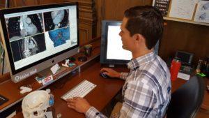
- Eric Myers working in Mimics

-

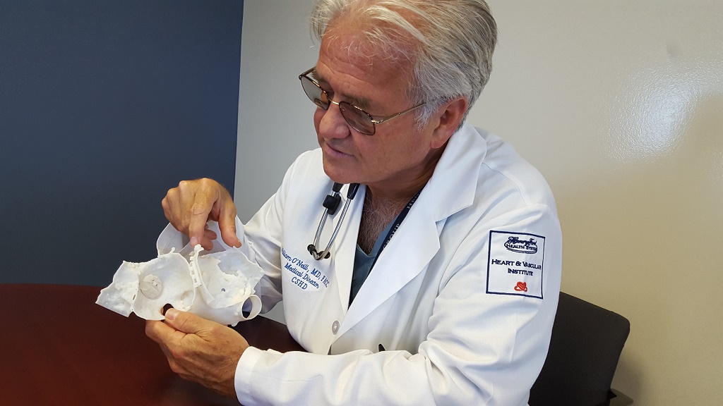
- Dr. William O’Neill examines a valve
-

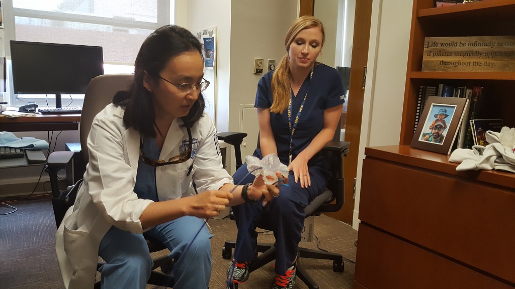
- Dr. Dee Dee Wang shows catheter placement while Marianne Rollet looks on
The Innovation Institute reflects its heritage as well as a strong push toward the future. While my eye was immediately drawn to Myers’ work station and the three 3D printers set up around it, across the room was the stage where Henry Ford used to hand out diplomas to newly graduated nurses. Sitting down with Myers, the conversation turned more toward the recent past.
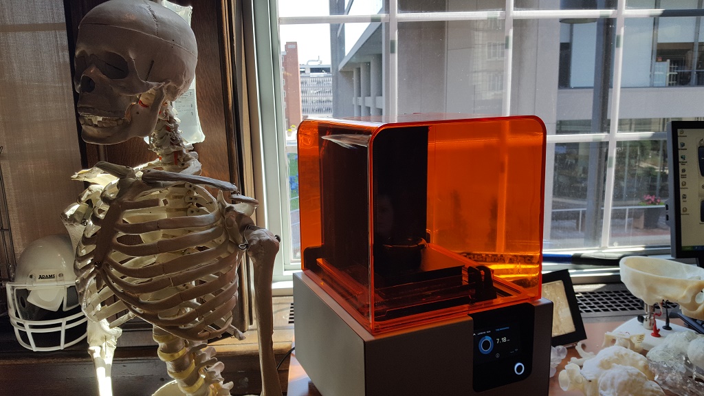
Getting started with 3D printing in this setting about two years ago, the team initially invested in “a little MakerBot” to start proofing out what might be possible in patient care. Moving on from the wooden-framed early MakerBot, the current FDM offering on-site is a Replicator 2X, which is now kept company as well by two higher-end machines. A Stratasys Objet30 Pro is the workhorse machine here, used for a large majority of current jobs, while a newer Formlabs Form 2 is offering new benefits with its wider portfolio of materials that might lead to what Myers described as exciting big next steps.


“The software is often overlooked in all this,” he added, motioning to his computer setup where the design work is done. “We’ve been using Mimics since the beginning, and have a great relationship with Materialise. I offered the CAD perspective, and had to learn the medical. We’ve been working closely with the cardiology team who are well-versed in imaging. It’s a really cool relationship.”
Relationships such as that with Materialise, whose software has been at the heart of the cardiac team’s imaging work, and with the cardiology department are driving this work forward. And of course relationships among the members of this team are paramount as a common goal unites them in bringing the technology into a hospital environment.
“Our passion is helping,” Dr. Wang said emphatically. “We have the perfect mix of people to get this started. It was Dr. O’Neill’s brainchild. He had the idea and tossed me the ball. There’s been lots of hard work from there, from Marianne and Eric, and support from administration and from philanthropic donors, from the Henry Ford Innovative Institute. We’re excited about it, it’s very right place, right time, right team. There’s a startup feeling, it’s very fast-paced, and a lot of fun. And a lot of hard work. Business, technology, patients — we’re bringing them all together, and this is the new healthcare.”

Dr. Wang and Rollet explain mitral valve procedures to me
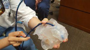
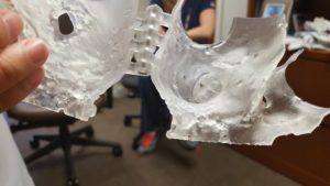
The work is ongoing and, indeed, fast-paced. While I talked with Myers, the Form 2 and Objet30 Pro were both hard at work, printing patient heart model number 521; in Dr. Wang’s office, she and Rollet were preparing for a procedure set to begin shortly. Dr. Wang demonstrated for me how a heart model Myers had 3D printed had already proven to be useful in proving out ahead of the procedure which of the three options of catheter would be the best fit for placing a device into the patient’s heart valve. Through this simple test ahead of time, the team cuts down on guesswork when it comes time for the actual procedure, reducing risks associated with switching out catheter sizes during the insertion process or of otherwise having an ill-fit tool.
“Each appendage is like a fingerprint,” Dr. O’Neill explained using another 3D printed heart model. “It’s totally different; we usually work with a 2D image, but this shows all the differences and how it interacts, allowing us to practice before the patient’s procedure. It’s great for safety. The time of the procedure is cut down and we have a success rate of about 99% now because we know the size, the location, the angulation; we can plan our entrance and approach. Our average procedure time is about 25 minutes, and we use one device per case on average. Other hospitals use two or three to try them out; we almost always get it right the first time. Pulling out risks a tear in the heart wall, but this reduces that chance.”

Dr. O’Neill demonstrates device placement within a 3D printed heart model
Key to the successes being seen now through the use of these 3D printed heart models is indeed that it is being seen now.
“This is not theoretical,” Dr. O’Neill told me. “We put this into action all the time. It is invaluable information that lets us plan, so we know ahead of time how the devices fit and where any potential problems will be. I’ve been practicing for about 30 years now; 3D printing is one of the biggest advances for structural heart.”
More than 500 patients’ lives have so far been touched by the use of advanced technology used at HFHS. Each of these lives has been impacted differently, and the goal for the hospital is to see this impact continue to spread, for 3D printing to become a mainstay in healthcare.
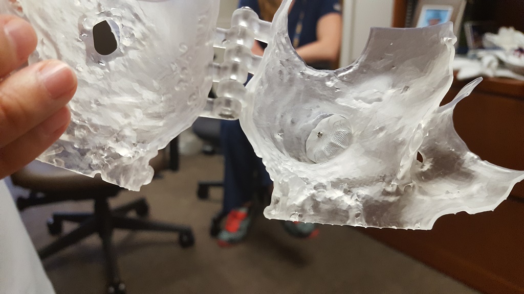
-
- This correctly sized/shaped piece will enter the heart here…

-
- …placing the device precisely here.
“There’s a massive amount of people we can help who we couldn’t help before,” Rollet told me.

Patents line the staircase in the Innovation Institute
Holding back further adoption is a gap between lab work and commercialization — as well as insurance companies’ being still a bit slow to approve 3D printing for reimbursement on their plans. Myers’ focus in the Innovation Institute is to commercialize IP from the team, getting it further into the world and into healthcare use. Regarding their work with 3D, Myers explained that the team has published around half a dozen papers and has worked already on three patents. Of those three, one has been granted, and licensed to Materialise, and the other two are applications. Beyond the all-important goal of touching patient lives, he noted, they have had some commercial success.
“There’s a huge gap between lab science and what’s available commercially,” Dr. Wang said. “Bridging that gap would be tremendous for patient care. That’s where the donations come in. There’s a constant need for patients, and showing how we’re using it to help them keeps the funding coming in. We’re incredibly fortunate to have the donors we do. Part of the funding we receive also goes to creating a 3D printed heart we send the patient home with. That means a lot to them, to hold their hearts.”

Dr. Wang and Rollet examine a heart model in front of the library of 3D printed hearts in Dr. Wang’s office
Spending the day with the team at HFHS was nothing short of inspiring. The technology developing around the world today can be put to use for any number of applications, but no vertical is more impactful to the human condition than healthcare. 3D printing is literally saving lives.
The work made possible through detailed medical imaging software and 3D printing at Henry Ford Hospital System is allowing for a new paradigm in healthcare. On top of the obvious benefits to patient care, arguably the biggest impact to be seen through this team’s work will come through their efforts towards commercialization, offering the potential to expand on the ability to care for patients whose hearts could use the help. We’ll be taking a further look at some of the information shared in coming days, as the work being done is extensive and potentially quite wide-reaching. Discuss in the Henry Ford forum at 3DPB.com.

Subscribe to Our Email Newsletter
Stay up-to-date on all the latest news from the 3D printing industry and receive information and offers from third party vendors.
Print Services
Upload your 3D Models and get them printed quickly and efficiently.


You May Also Like
AM Asia Watch: China’s HeyGears Lands $44M to Expand Beyond Dental 3D Printing
Chinese 3D printing company HeyGears raised more than 300 million Yuan (roughly $44 million) in a new Series C funding round as it looks to expand beyond its industrial and...
The University of Utrecht: “3D Printing Could Change Who Gets to Become a Manufacturing Power”
For decades, manufacturing has mostly been controlled by countries with huge factories, lower labor costs, and industrial systems that took years, sometimes decades, to build. But Utrecht University human geographers...
3D Printing News Briefs, May 28, 2026: Continuous Fiber Reinforcement, Bioprinted Trachea, & More
In today’s 3D Printing News Briefs, America Makes announced the winners of its JAQS-SQ Project Call. Axtra3D is partnering with Keystone Industries to expand its dental material ecosystem, while BigRep...
Asia AM Watch: China’s SHINING 3D Restarts IPO Review Process
SHINING 3D is moving forward again with its plans to go public in China, after restarting its Beijing Stock Exchange (BSE) initial public offering (IPO) review process and filing updated...