PhD Candidate Turns to 3D Hubs to Create Realistic, Functioning 3D Printed Neonatal Models for Medical Training

![]() It’s hard when a loved one has to undergo surgery. Obviously, it’s difficult for the patient, but it’s also hard to sit in the waiting room, powerless, hoping the doctor comes in soon, but not too soon, and gives you good news. I don’t have personal experience with this, but I imagine it’s probably even worse when a newborn has to have surgery – they’re so tiny, and fragile. What’s even scarier about neonatal surgery is the lack of complex, accurate medical models. It’s very helpful for nurses and surgeons alike to interact and practice with realistic anatomical models before medical procedures and surgeries – it can really increase the rate of success. Medical models are also good training tools for medical students. Most neonatal practice mannequins are basically just dolls equipped with a spring, and don’t feel anything like a real newborn. But PhD candidate Mark Thielen, from the Technical University of Eindhoven (TU/e) in the Netherlands, recently worked with online 3D printing service platform 3D Hubs to change that.
It’s hard when a loved one has to undergo surgery. Obviously, it’s difficult for the patient, but it’s also hard to sit in the waiting room, powerless, hoping the doctor comes in soon, but not too soon, and gives you good news. I don’t have personal experience with this, but I imagine it’s probably even worse when a newborn has to have surgery – they’re so tiny, and fragile. What’s even scarier about neonatal surgery is the lack of complex, accurate medical models. It’s very helpful for nurses and surgeons alike to interact and practice with realistic anatomical models before medical procedures and surgeries – it can really increase the rate of success. Medical models are also good training tools for medical students. Most neonatal practice mannequins are basically just dolls equipped with a spring, and don’t feel anything like a real newborn. But PhD candidate Mark Thielen, from the Technical University of Eindhoven (TU/e) in the Netherlands, recently worked with online 3D printing service platform 3D Hubs to change that.
TU/e is certainly no stranger to 3D printing – we have discussed their work with concrete 3D printing before. But Thielen’s amazing work is about as far away from concrete as you can imagine: he was able to create functional, lifelike internal structures and organs for neonatal training models using MRI scans and 3D printing. His new training models, which even feature embedded sensors, have the potential to save countless newborn lives, and would not be possible to create through traditional production technologies.

PhD candidate Mark Thielen with his 3D prints, provided both by 3D Hubs and his internal printers.
Thielen, a Healthcare Flagship Program participant, is working hard to increase the success of medical procedures and surgeries for neonatal patients. His research centers around developing realistic training mannequins, complete with functioning internal organs and sensors that can monitor stress, pressure, and impact during trial procedures, like intubation and CPR, and provide intelligent feedback. By using the 3D Hubs network, he had access to a range of materials, and was able to produce the necessary parts locally. The neonatal training mannequins have two important components: the combination of the rib cage and spine, which together house the second component, the internal organs. 3D printing allowed Thielen to realistically produce newborn anatomy, thanks to the organic shapes the technology can create, along with decreased lead time and costs.
-
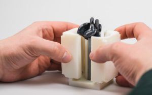
- PolyJet mold for the creation of the heart using flexible material as support (black) and rigid plastic as the surround (white).

-
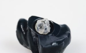
- Silicon heart created from a 3D Printed mold, with functioning valve (PolyJet)

-

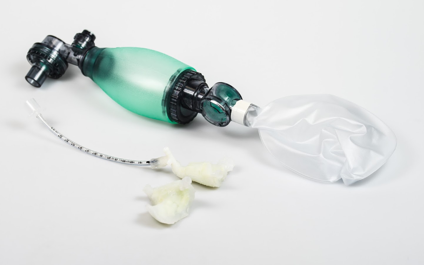
- Functioning silicon lungs made with PolyJet mold, with air pump for simulation
There are five steps Thielen completed to create his models:
- An MRI scan of a newborn patient is taken
- Materialise Mimics software segments specific organs in the scan and reconstructs them into 3D printable files
- The files are imported into SOLIDWORKS
- Materialise Magics software is used to ensure the organ models are 3D printed smoothly
- The files are exported to a 3D printer
Initial tests were completed using different thermoplastic elastomers on a desktop FDM 3D printer, which was also used to make the larger parts of the model, like the rib cage. Once the designs were ready, SLS 3D printing was used to create the final rib cage, due to its dimensional freedom and accuracy. The molds for the functional organ models were 3D printed using PolyJet technology.
 3D printing was really the best way for Thielen to go for this research project. Molds that are created using 3D printing, compared to using conventional manufacturing methods, give users the freedom to make rapid design changes, and PolyJet printing allows materials to be combined, like both flexible and rigid plastics. For example, neonatal heart models are extremely small, but need to show high levels of detail, due to the organ’s working valves. So really, 3D printing was the only way to go when creating molds for these parts.

Compression testing to check how much force the ribcage can take and how it affects the internal organs and flow of blood (liquid) around the model
Once 3D printing is complete, and the models have been removed from their molds, Thielen combines the rib cage, spine, and internal organs, all of which function realistically: the lungs even inflate and deflate. He next installs cameras and sensors, which provide feedback during different trial procedures, and runs a fluid, which models blood, through the mannequin. This liquid works in tandem with the sensors and cameras and monitors the flow through the mannequin. It will essentially act like a signal when test pressure is too little, or too high.
Thielen’s inspiring research into creating realistic mannequins is incredibly important to the success rate of neonatal surgeries, giving nurses and physicians valuable and necessary training opportunities. But, he believes his work could have wider applications than just the field of neonatal medicine.
Checking the neonatal MRI scans, to export the relevant structures and prepare for 3D printing
“I believe that developing and advancing what we started here can aid medical research in a broader scope,” explained Thielen. “We could potentially create realistic patient models of other body parts to strengthen medical training for emergency procedures and pregnancies.”

To get a better glimpse of Thielen’s models, and learn about some of the tests they undergo, check out this short video (note, there is no sound):
Discuss in the Neonatal Models forum at 3DPB.com.
[Source/Images: 3D Hubs]
Subscribe to Our Email Newsletter
Stay up-to-date on all the latest news from the 3D printing industry and receive information and offers from third party vendors.
Print Services
Upload your 3D Models and get them printed quickly and efficiently.


You May Also Like
AM Asia Watch: China’s HeyGears Lands $44M to Expand Beyond Dental 3D Printing
Chinese 3D printing company HeyGears raised more than 300 million Yuan (roughly $44 million) in a new Series C funding round as it looks to expand beyond its industrial and...
The University of Utrecht: “3D Printing Could Change Who Gets to Become a Manufacturing Power”
For decades, manufacturing has mostly been controlled by countries with huge factories, lower labor costs, and industrial systems that took years, sometimes decades, to build. But Utrecht University human geographers...
3D Printing News Briefs, May 28, 2026: Continuous Fiber Reinforcement, Bioprinted Trachea, & More
In today’s 3D Printing News Briefs, America Makes announced the winners of its JAQS-SQ Project Call. Axtra3D is partnering with Keystone Industries to expand its dental material ecosystem, while BigRep...
Asia AM Watch: China’s SHINING 3D Restarts IPO Review Process
SHINING 3D is moving forward again with its plans to go public in China, after restarting its Beijing Stock Exchange (BSE) initial public offering (IPO) review process and filing updated...