MIT & Boston Children’s Hospital Conducting Study to See if 3D Printed Heart Models Offer Improvement in Surgical Outcome


3D printing offers enormous reward in that you can make things pretty much however you deem fit–it’s your 3D design and you can make it or tweak it at whim, enjoying the latitude afforded by scalability and customization.
For items like apparel, accessories, and on up to larger items like high-quality industrial components for aerospace and the automotive sectors, it almost seems to be too good to be true when compared to the limits of traditional processes. But when examined in terms of what can be done within the medical arena, the implications are stunning for improving–and often saving–human lives.
![]() While we often write about medical models being created through 3D printing, an important new research program is being created in regards to 3D printed heart models created through MRIs.
While we often write about medical models being created through 3D printing, an important new research program is being created in regards to 3D printed heart models created through MRIs.
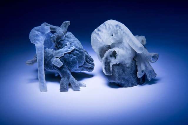
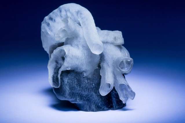
Researchers at MIT and Boston Children’s Hospital have recently created a way to create the 3D printed models in a matter of hours, and their innovation will be undergoing a study this fall to see just how useful these medical models of the heart are.
Seven surgeons will be participating in the study, using MRIs from ten different patients as they gauge the effectiveness of the 3D prints which can be used not only in education but most importantly in diagnosing conditions–as well as assisting in surgeries.
“Our collaborators are convinced that this will make a difference,” says Polina Golland, a professor of electrical engineering and computer science at MIT, who led the project. “The phrase I heard is that ‘surgeons see with their hands,’ that the perception is in the touch.”
While currently it is not that unusual to see MRIs being converted for 3D printed medical models, the study is meant to take a deeper look at whether this process is valid–and viable–in terms of clarity for heart surgery. There are concerns regarding the variety of shades in the MRI data and whether or not edges are always depicted clearly. This is a common issue, however, and it’s referred to as ‘image segmentation.’ Because it’s generally not possible to get the accuracy required digitally, the researchers at hand have been hard at work developing the new system which was funded by the Boston Children’s Hospital as well as Harvard Catalyst, a consortium which places a strong emphasis on scientific innovation.
Using a model of a heart for diagnosing and surgery is risky in that many patients have irregular patterns due to their conditions. If everything is not precise within the scan and corresponding 3D model, a life could be put at risk when it is used for diagnosis and surgery. Currently, in terms of gray area, medical professionals fix this problem themselves by manually indicating boundaries in MRI scans–but with hundreds of cross sections to deal with, this is a job that could take a full day’s work.
“They want to bring the kids in for scanning and spend probably a day or two doing planning of how exactly they’re going to operate,” Golland says. “If it takes another day just to process the images, it becomes unwieldy.”
 What they are doing currently, is quite simply, trying to make models that clearly show ‘idiosyncracies’ which are common in heart conditions and must be accurately portrayed in a model. While the algorithms they generally use are not usually accurate enough, Golland and her team worked with an expert so that a new and much more efficient algorithm could be created. They marveled at the results after they segmented just 14 patches, letting the algorithm infer the rest–and yielding 90 percent agreement with expert segmentation of the entire collection of 200 cross sections.
“I think that if somebody told me that I could segment the whole heart from eight slices out of 200, I would not have believed them,” Golland says. “It was a surprise to us.”
The surgeons will be given data with scans, 3D models, and they will also draw up plans for surgery to be compared with documentation regarding what was performed on patients. With all of this information they hope to ascertain whether or not the 3D models are actually an improvement on traditional practices.
“Absolutely, a 3-D model would indeed help,” says Sitaram Emani, a cardiac surgeon at Boston Children’s Hospital who is not a co-author on the new paper. “We have used this type of model in a few patients, and in fact performed ‘virtual surgery’ on the heart to simulate real conditions. Doing this really helped with the real surgery in terms of reducing the amount of time spent examining the heart and performing the repair.
“I think having this will also reduce the incidence of residual lesions — imperfections in repair — by allowing us to simulate and plan the size and shape of patches to be used,” Emani adds. “Ultimately, 3D-printed patches based upon the model will allow us to tailor prosthesis to patient.
“Finally, having this immensely simplifies discussions with families, who find the anatomy confusing,” Emani says. “This gives them a better visual, and many patients and families have commented on how this empowers them to understand their condition better.”
 The research and work being performed will be presented in detail at the International Conference on Medical Image Computing and Computer Assisted Intervention being held in October. Both Golland and her colleagues will describe their new system.
What do you think? Is the surgical outcome improved through the use of 3D printed heart models? Discuss in the 3D Printed Heart forum thread on 3DPB.com.
Danielle Pace, an MIT graduate student in electrical engineering and computer science, is first author on the paper and spearheaded the development of the software that analyzes the MRI scans. Medhi Moghari, a physicist at Boston Children’s Hospital, developed new procedures that increase the precision of MRI scans tenfold, and Andrew Powell, a cardiologist at the hospital, leads the project’s clinical work.
[Photos: Bryce Vickmark]Subscribe to Our Email Newsletter
Stay up-to-date on all the latest news from the 3D printing industry and receive information and offers from third party vendors.
Print Services
Upload your 3D Models and get them printed quickly and efficiently.


You May Also Like
Scientists Create Stretchy 3D Printed Implants for High Blood Pressure Treatment
Researchers at Pennsylvania State University (Penn State) say they may have found a softer, less invasive way to treat severe high blood pressure. In a new study published in the...
Harvard’s Jennifer Lewis Lab Is 3D Printing Artificial Muscles That Twist and Bend on Demand
Researchers at Harvard John A. Paulson School of Engineering and Applied Sciences (SEAS) have developed a new way to 3D print materials that can move on their own, bending, twisting,...
3D Printing News Briefs, May 2, 2026: Soft Robots, Agricultural Waste, & More
In this weekend’s 3D Printing News Briefs, we’ll start off with a multi-laser metal powder bed fusion 3D printer and post-processing news. We’ll end with research into soft robotics and...
Harvard SEAS Engineers Develop 3D Printing Method for Soft Robotic Components with Programmable Shapes
The world of soft robotics is still largely in its pure research phase, but the R&D landscape has started to produce examples of early-stage commercialization. Researchers have started to refine...