
Breakthrough 3D Printed Video Microscope Might Help Defeat the African Eye Worm

Lymphatic filariasis might be defeated with this cellphone-based, 3D printed microscope and diagnostic device.
Lymphatic filariasis is a nasty condition caused by the parasitic African Eye Worm, a microscopic thready creature which, as an adult, can can only live within the human lymph system. The condition affects more than 120 million people in the tropics and sub-tropics of Africa, Asia and parts of Central and South America.
It spreads from person to person via mosquito bites, and as a mosquito bites a person who has lymphatic filariasis, the worms circulating in that person’s blood enter – and infect – the mosquito. As the infected mosquito bites the next person, the tiny worms pass from the mosquito through a human’s skin and travel to the lymph vessels where they can grow into adults.
The adult worm lives for some 7 years, and as they mate, they can release millions more of their microscopic progeny into the blood.
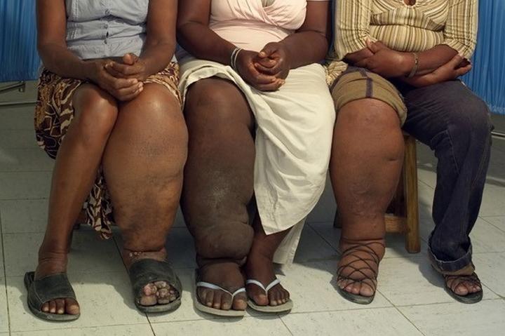
 Most people are completely unaware that they have lymphatic filariasis – at least initially. Victims don’t feel symptoms until the adult worms die, and while the condition isn’t usually life threatening, it can cause horrific, permanent damage to the lymph system and kidneys. That damage ultimately causes fluid to collect and causes swelling in the arms, breasts, legs and even the genital area.
It’s bad news and the entire leg, arm or genital area may swell to several times normal size. The diminished function of the lymph system makes it difficult for the body to fight off germs and infections and it can also cause hardening and thickening of the skin, which is called elephantiasis. It’s a leading cause of permanent and long-term disability in sufferers around the world.
Now a research team of UC Berkeley engineers have used 3D printing to develop a smartphone microscope which can detect and quantify the infection of parasitic worms in a single drop of blood.
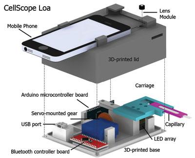
They call it the Cellscope Loa, and it promises a simple to use solution to a problem which has tormented sufferers and health workers.
“We previously showed that mobile phones can be used for microscopy, but this is the first device that combines the imaging technology with hardware and software automation to create a complete diagnostic solution,” says Daniel Fletcher, an associate chair and professor of bioengineering at UC Berkeley. “The video CellScope provides accurate, fast results that enable health workers to make potentially life-saving treatment decisions in the field.”
Dr. Thomas Nutman of the National Institute of Allergy and Infectious Diseases collaborated with the UC Berkeley team and workers from Cameroon and France to develop the Loa Cellscope, and commence a pilot study in Cameroon.
The CellScope Loa uses motion detection rather than molecular information or fluorescent staining to detect the movement of the Eye Worm, and the researchers say it’s every bit as accurate as the conventional screening method. The results of the study were published in the journal Science Translational Medicine.
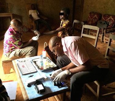
Tests on 40,000 patients in Cameroon are underway now of the Cellscope Loa.
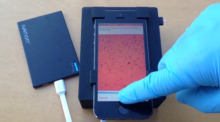
A 3D printed holder containing a high resolution lens is located under the phone’s camera, and an Arduino circuit board and Bluetooth connection take care of business. A blood sample loaded into the case is subjected to review by complex algorithms to detect the wriggling motion which characterizes the presence of the eye worms.
The device then shows a count on the phone to let doctors know if the treatment might be dangerous to the patient. Treatment via a drug, Ivermectin of Stromectol, is very risky for patients with high levels of Loa worms in their systems.
The test itself takes under three minutes, and as the process is completely automated, very little training is required to effectively use the device.
Fletcher says the technology could well be adapted to for a variety of other diseases such as malaria and TB.
Are you aware of any other projects which use smartphone technology to solve problems in health or scientific issues? Let us know in the 3D Printed Microscope And Diagnostic Device forum thread on 3DPB.com.
Subscribe to Our Email Newsletter
Stay up-to-date on all the latest news from the 3D printing industry and receive information and offers from third party vendors.
You May Also Like
3D Printed Heat Spreader Could Improve Efficiency of Electronics
The low-hanging fruit for decarbonization has long been improving the efficiency of existing systems, hence the justification for LED lights and ENERGY STAR certified appliances. While such minor moves are...
3D Printing News Unpeeled: Marine Gearboxes, 3D Printed Motors and $1.7 Million in Seed Funding
UK based Equipmake just released their Ampere-220 e-axle system. The system, which is meant for high performance electric cars, was similar to one released on the Ariel HIPERCAR. It has...
CEAD Unveils 36-Meter-Long 3D Printer for Abu Dhabi’s Al Seer Marine
CEAD, a Dutch original equipment manufacturer dedicated to large-format 3D printers, has unveiled what it claims to be the world’s largest robotic arm-based 3D printer. At 36 meters long and...
3D Printed Biocomposites Could Help Reduce Marine Plastic Pollution
Concerns about the impact of plastic litter and microplastics in the oceans are at the forefront of environmental study. For decades, the marine environment has suffered from the degradation of...