Drug Response Testing: Bioprinting Scaffold-Free Cardiac Constructs

Researchers have come together to make improvements in cardiac medicine, releasing the details of a recent study in “Drug response analysis for scaffold-free cardiac constructs fabricated using bio-3D printer.” Acting on previous research which resulted in bioprinted tubular cardiac constructs, the scientists have now continued their work to create a technique for evaluating contractile force and drug response.
While striving to eliminate the need for animal testing or clinical studies, the researchers realize the ongoing requirement to measure the effects of drugs on humans, stating, “understanding the drug response of the heart is essential for new drug development.”
In many cases, testing requires substantial time and expense and can also be detrimental to the health of those participating in studies. Developing cardiac constructs without scaffolds allowed for comprehensive and accurate evaluation of drug response.

Bio-3D printing process and drug response of cardiac constructs. Cells aggregate as spheroids. The appropriate needle array and desired 3D design were selected and prepared (a). The spheroids were then printed onto the needle array (b). After cultivation, the drug response was measured using the fabricated cardiac constructs (c).
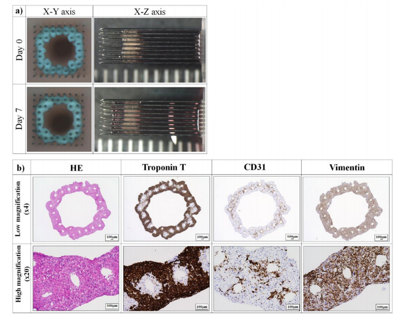
Fabrication of scaffold-free tubular cardiac constructs. (a) Representative images of the fabricated tubular cardiac constructs immediately following printing and culture on the needle array for 7 days. (b) Immunohistochemical analysis of the cardiac constructs. The samples were observed at low (4×) and high (20×) magnification. iCells were identified by troponin T staining, whereas HUVECs and NHDFs were stained with CD31 and vimentin, respectively. Scale bar = 100 μm.
Upon creating a contraction analysis system, the authors tested:
- Electrical stimulation
- Temperature dependence of spontaneous contraction in cardiac constructs
- Drug reactivity analysis of cardiac constructs
- Cytotoxicity effect of doxorubicin on cardiac constructs
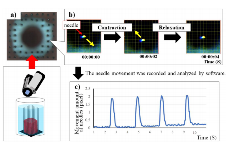
Motion analysis of the needle array movement by contraction of cardiac constructs. The cardiac constructs on the needle array were recorded (a), and the movement of the needle array was tracked using sofware (b). Measurement and analysis of the needle movements (c).

Change in the spontaneous contractile frequency and force of cardiac constructs in culture. Four points on the needle arrays were selected to evaluate contraction of the cardiac construct (a), and the changes in beating rate/ 10 s (b) and in the contractile frequency and force of cardiac constructs (c) were analyzed at day 0 and day 7. N=8 for each group, *P<0.01. Error bars represent standard deviation. The movements of the needle array of the cardiac constructs at day 0 and day 7 were compared (d,e).
To monitor the ‘beating rate’ of the cardiac constructs, the researchers established continued assessment of electrical stimulation:
“During the spontaneous contraction condition, the beating rate of cardiac constructs was 3 beats per 10s. When 1 or 2 Hz-paced electrical stimulation was applied to cardiac constructs, the beating rate increased. However, 2Hz paced electrical stimulation prevented full relaxation of the cardiac constructs and showed a decrease in the top movement of the needle. Stevens et al. also demonstrated that scaffold-free cardiac patches showed a decrease in contractile force at 2Hz or 3Hz paced electrical stimulation26. Thus, our results indicate that this contraction analysis system can reproduce the contraction of the human heart.”
Consistency in temperature is required for the cardiac constructs, with variations affecting the beating rate—leaving the researchers to note that such control is a ‘very important parameter’ in such testing. Moving forward in the study, four drugs were used during experimentation: isoproterenol, propranolol, blebbistatin, and doxorubicin.
“We selected well-known drugs, isoproterenol and propranolol, because of their ability to change the beating rate and contractile force of cardiomyocytes,” said the authors.
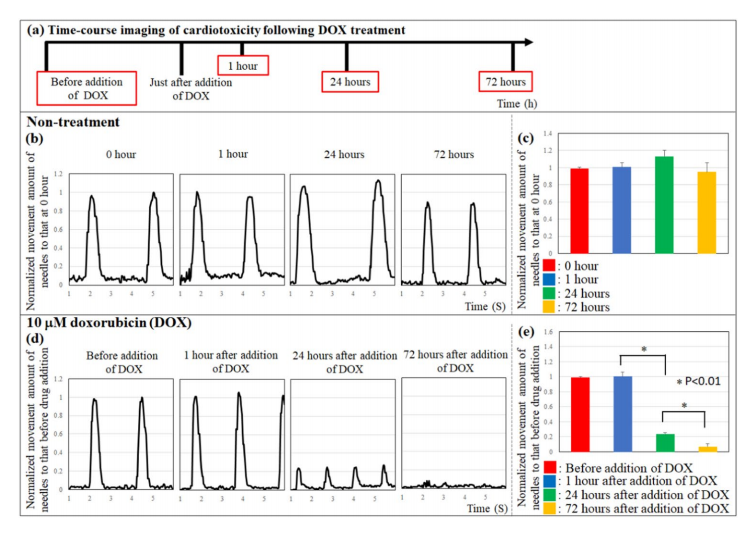
Cardiotoxicity in cardiac constructs following doxorubicin treatment. Time-course imaging of cardiotoxicity following DOX treatment (a). Te movement of the needle array did not change in response to non-treatment (b,c). Conversely, the top movement of the needle array decreased in response to 10 μM doxorubicin treatment (d,e). N=3 for each group, *P<0.01. Error bars represent standard deviation.
Because evaluating the dangers of cardiotoxicity was critical in the study, the researchers focused on doxorubicin (DOX) as a ‘model drug’ for such an assessment. The results were in line with other studies, indicating that the beating rate did not vary at one hour after the application of DOX. Overall, all factors showed that cardiotoxicity could be monitored with the new system and follow-up analysis.
“We could not calculate the contractile force of the construct, because the length of needle array was not uniform at the micro-level,” concluded the researchers. “In future, we will evaluate the contractile force accurately by improving the length of the needle array. Second, we used the frame rate (30 frames per s) of our video camera to evaluate contraction of the cardiac constructs. Conversely, Takeda et al. reported that a video camera recording at 150 frames per s can measure the contraction velocity of cardiomyocytes as well as the relaxation velocity and contraction-relaxation velocity. Tus, in the future, we aim to perform contraction analysis of cardiac constructs using a video camera with a high frame rate.
“This analysis method can be used for new drug development, due to its high drug response predictability in vitro. We believe that accuracy of this contraction analysis system will increase in the future due to advances in camera technology and the quality of the needle array.”
Scientists, manufacturers, engineers, and a variety of users around the globe continue to shed constraints that cause challenge or extra steps in digital fabrication—from industrial applications in aerospace, automotive, and construction, to bioprinting, where refining the process could be critical to the ultimate goal of being able to fabricate amazing items like human organs
What do you think of this news? Let us know your thoughts! Join the discussion of this and other 3D printing topics at 3DPrintBoard.com.
[Source / Images: ‘Drug response analysis for scaffold-free cardiac constructs fabricated using bio-3D printer’]Subscribe to Our Email Newsletter
Stay up-to-date on all the latest news from the 3D printing industry and receive information and offers from third party vendors.
You May Also Like
Air Force Awards Fortius Metals $1.25M to Qualify 3D Printing Wire for Hypersonic Applications
AFWERX, part of the US Air Force Research Laboratory (AFRL), awarded a Direct-to-Phase II Small Business Innovation Research (SBIR) contract worth $1.25 million to Colorado’s Fortius Metals, to accelerate qualification...
US Air Force Awards JuggerBot $4M for Large-format Hybrid 3D Printing
Large-format 3D printer manufacturer JuggerBot has received a $4 million grant to develop a large format 3D printer, courtesy of the Under Secretary of Defense, Research and Engineering Manufacturing Technology...
Where Have All AM’s Unicorns Gone?
In the rapidly evolving world of 3D printing, startups valued at over a billion dollars, known as unicorns, once seemed as fantastical as the mythical creatures themselves. While a few...
How My Childhood Fascination with Planes Led to Investing in 3D Printing
My fascination with aerospace started young, and I started studying planes–identifying them in the sky and learning everything I could about how they work. Fast forward to my first week...