
3D Printing, VR & MR for Better Lung Surgery Outcomes


Scientists from Guangdong Academy of Medical Sciences have released the findings of their recent study in ‘Three-dimensional Printing, Virtual Reality and Mixed Reality for Pulmonary Atresia: Early Surgical Outcomes Q1 Evaluation.’
Delving into the realm of cardiac medicine and associated 3D-printed models, the authors were able to use both virtual reality (VR) and mixed reality (MR) for better understanding of anatomy and surgical planning in cases dealing with a congenital heart disease: pulmonary atresia (PA) with ventricular septal defect (VSD) and major aortopulmonary collateral arteries (MAPCA). Surgical management can be challenging, and despite some success in both short- and long-term outcomes, “prognosis is still poor.”
Typical exploration of such conditions, using CTs and MRIs, as well as catheter angiography does not always yield the comprehensive knowledge required for treatment.
“Recent studies have shown similarities between the 3D heart model and real cardiac anatomy and suggested a more systematic approach to explore the correlation between the 3D heart and actual anatomical details on plain CT scans or MRI,” stated the researchers.
“With the aid of holographic visualization, traditional images are converted into 3D models with detailed information and excellent image quality. Previous studies have evaluated either optimization of the surgical strategy or the impact on surgical outcomes. However, few studies have reported the combination of 3D printing, VR and MR in patients with PA/ VSD/MAPCA and their role in guiding medical treatment.”
Patients’ basic clinical characteristics
Scans were completed for all patients participating in the study, with 3D images made for VR “by one projection per eye.” This allowed for a suitable sensation of depth, allowing for viewing in “a virtual world separated from the real surrounding.” Models were 3D printed at a 1:1 scale and measured by an expert. Surgeons were then able to perform surgical simulation using 3D glasses.
“The true 3D depth horizon from VR was integrated with the real-world environment during surgery,” explained the researchers.
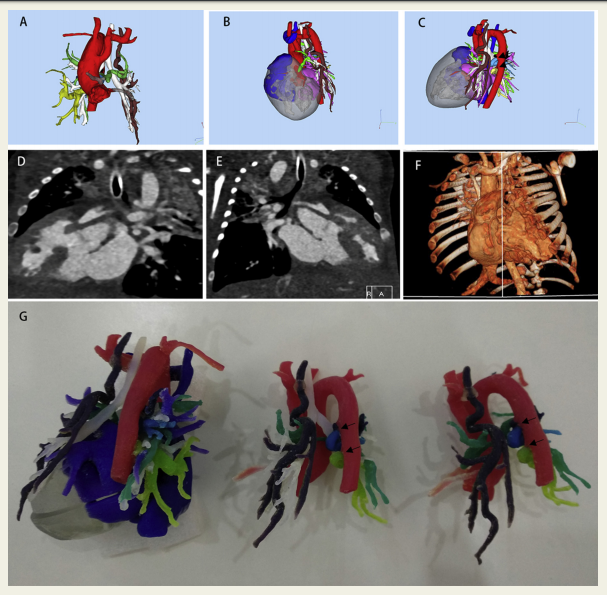
Selected computed tomography (CT) angiography and complete preoperative models in a patient with pulmonary atresia and major aortopulmonary collateral arteries (MAPCA) (Case 1). A–C. Data and images derived from CT scanning. D–F. Radiological image and segmentation. The collateral vessels were obscure in CT images. G. Final three-dimensional printed model. *Arrows in C and F indicate MAPCA
Following procedures, all patients in the study reflected “satisfactory postoperative recovery” while in the hospital; later, however, three displayed complications showing a right bundle branch block and ST segment change, one requiring chest drainage, and another suffered from pneumonia. Another patient was found to have an arrhythmia.
Ultimately, the research showed that all new technologies used in the study “enhanced the surgeons” understanding of anatomy, with “acceptable” outcomes.
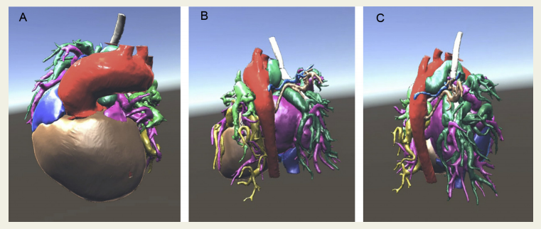
Virtual reality hologram of pulmonary atresia/ventricular septal defect/major aortopulmonary collateral arteries (Case 2). *Arrows in B and C indicate major aortopulmonary collateral arteries
“There were several advantages of 3D printing applications in patients with PA/VSD/MAPCA in this study. First, evaluation of the 3D printing allowed the surgical teams to preoperatively analyze their plans. In previous studies, surgical decisions varied from physiological palliation to biventricular repair after evaluating the 3D heart models. The current study failed to observe similar variations, which was probably due to the small sample size,” concluded the researchers.
“Second, optimal surgical outcomes depend on a thorough understanding of the anatomical structures in these PA cases. A thorough understanding is essential for surgeons to preclude unexpected findings and effectively limit the duration of the intervention. Moreover, surgeons can share their interpretations and views during surgery, with the help of surgical simulation using 3D models. Cardiovascular 3D models play an indispensable role in educating the parents, particularly during the decision-making and consent-signing processes.”

What do you think of this news? Let us know your thoughts; join the discussion of this and other 3D printing topics at 3DPrintBoard.com.
[Source / Images: ‘Three-dimensional Printing, Virtual Reality and Mixed Reality for Pulmonary Atresia: Early Surgical Outcomes Q1 Evaluation’]Subscribe to Our Email Newsletter
Stay up-to-date on all the latest news from the 3D printing industry and receive information and offers from third party vendors.
Print Services
Upload your 3D Models and get them printed quickly and efficiently.


You May Also Like
Additive Manufacturing at a Crossroads
Additive manufacturing is at a crossroads. Simultaneously, we find ourselves between certain very different modalities, applications, and industries. Rather than being able to explore them all, companies will now have...
After 17 Years at 3D Systems, Katie Weimer Is Betting on Regenerative Breast Tissue
After spending 17 years helping build healthcare applications at 3D Systems and its predecessor Medical Modeling, Katie Weimer wasn’t planning to launch a startup. But when a regenerative breast tissue...
Why Elegoo Chose Emoji® to Introduce More People to 3D Printing
When Elegoo unveiled the world’s first officially licensed emoji®-themed 3D printer, it wasn’t just launching another version of an existing machine. The company was testing a much bigger idea by...
The Longevity Economy Needs a Factory
Longevity has become one of the biggest stories in healthcare. Every week seems to add a new announcement about an anti-aging therapy, an AI-powered drug discovery platform, or a startup...


