
Plastic Surgery: 3D Printed Patient-Specific Applications for Craniomaxillofacial Operations


Plastic surgery can help improve quality of life, especially when patients are dealing with craniofacial issues – soft tissues of the face, like the nose and lips, and bones of the craniomaxillofacial (CMF) skeleton. This field helps improve those things that make us human, such as how we talk, look, and use our hands. 3D printing is often used in conjunction with plastic surgery, and, as a pair of researchers in Missouri explain, it’s an excellent specialty in which to better define how the technology is used in surgery.
Alexander Y. Lin, MD and Lauren M. Yarholar, MD, with the Saint Louis University School of Medicine and SSM Health Cardinal Glennon Children’s Hospital, published a paper, “Plastic Surgery Innovation with 3D Printing for Craniomaxillofacial Operations,” about the four types of 3D printed objects for surgical use, emphasizing plastic surgery and CMF operations, and the technology’s “high clinical relevance to all potential procedures utilizing 3DP assistance.”
“Plastic Surgery restores unique human qualities such as appearance, speech (palate), hands, to improve interaction with others and quality of life,” the two wrote. “Three-dimensional printing technology can be applied to Plastic Surgery craniomaxillofacial operations to change the bony skeleton of the skull, face, and jaws.”
The academic pediatric hospital where Dr. Lin and Dr. Yarholar work adopted in-house 3D printing in 2017, which has resulted in “faster turn-around, immediate customization, and decreased individual case costs after original investment.” Their group recently published a review with “a taxonomic analysis of patient-specific 3D printed models” used in CMF operations. The review only included patient-specific intraoperative 3D printing applications, and analyzed current research by finding and pulling data about this topic from peer-reviewed journal articles.
“We then concentrated on the specific clinical applications of craniomaxillofacial surgeries, resulting in 335 patients who had 3D-printed patient-specific data that was actually translated for use in surgery,” they wrote. “We analyzed these craniomaxillofacial operations to find patterns in the usage of 3DP for direct patient-specific surgical application, and discovered four main categories of utilizing 3DP for patient-specific craniomaxillofacial surgeries.”

Applications for Patient-Specific 3D Printing in Craniomaxillofacial Operations
These categories were further classified into four types of 3D printed patient-specific applications:
- Type I: contour models
- Type II: guides
- Type III: splints
- Type IV: implants
The most straightforward of these medical applications, 3D printed contour models are made from a patient’s imaging data. They can be used in nearly any surgical procedure that requires additional visualization and precision, and to show a patient’s hidden anatomy – very useful for fixing pediatric facial fractures, as these require precise fragment reduction, small incisions, and quick turnaround.

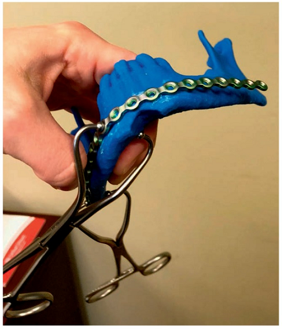
Figure 1: The white model is the patient’s injured mandible from a gunshot wound. The blue model was made by taking the less injured left side of the white mandible, and mirroring it to create a theoretically perfect mandible. The maxilla is printed as the blue skull to show that the theoretical blue mandible’s condyles were seated within the TMJ joints, indicating it has normal size and shape.
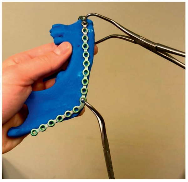
This demonstrates the reconstruction plate being pre-bent to a 3D printed predicted pre-injury model.
The figures above are models the researchers fabricated on their in-house 3D printer for a patient with a comminuted mandible fracture from a gunshot wound. The patient-specific mandible model was used to pre-bend reconstruction plates, which were brought into the OR to help “internally fixate the mandible fracture.” This made the fracture repair “faster and more precise.”
“The 3D-printed mandible (white) in figure 1, is the patient’s mandible after the gunshot injury – note the free-floating butterfly segment with multiple smaller fragments. Using our software, the uninjured image of the (left) side of the mandible was used to make a mirror image to replace the injured fractured (right) mandible,” the pair explained.
Guides are a little harder to make, because the negative space must be “mathematically characterized” in order for it to conform to a surface, like bone; this requires more precise and complex computer and image processing. 3D printed guides are often used in pediatric craniofacial reconstruction, such as irregular bone defects found in the skull. Finding an area of the skull for a donor site, making an irregular donor to match the defect, and finding that it’s not a perfect fit wastes a lot of time. 3D printed skull defect guides save time and bleeding morbidity for these young patients.
-

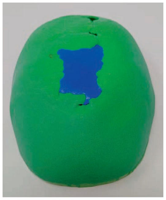
- Left, aerial view of patient’s cranium with skull defect, with the 3DP guide fitting in the cranial defect. This blue 3DP guide can be used during surgery to harvest a precisely shaped bone graft.
-
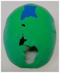
- Right, posterior to anterior view of the cranium and cranial defects with a 3DP guide in one of the two large cranial defects. A separate 3DP guide was used for this second, distinct skull defect.

The figures above “show a patient’s skull with a large midline cranial skull defect,” which was 3D printed, along with the blue guide; the latter was made with a resin that’s able to hold up under deformation temperatures during sterilization.
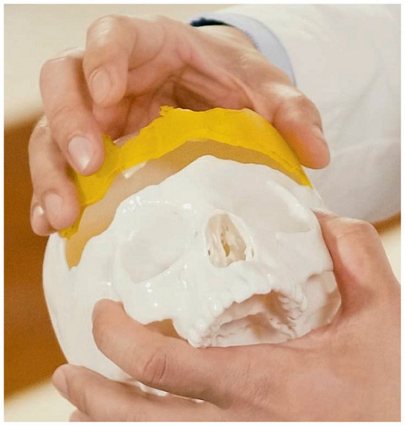
Patient with complex fronto-orbital craniofacial defect, with a 3DP yellow guide. The guide captures the unpredictable shape of the defect and the complex 3D angulation of the lateral aspects of the forehead where it sweeps into the temple region. This guide allows harvest of bone that has this congruent angle, ensuring an autologous bone graft that matches the patient’s forehead contour.
Even more computing and engineering are required to 3D print splints, as these need to “accurately perform virtual surgery to virtually cut the anatomy and move the pieces to the final position.” These are commonly used for jaw surgeries, especially for fixing under and overbites in children 16-18 years who have reached skeletal maturity, and children born with cleft lips that have a midface smaller than their lower jaw.
“Before 3DP splints, custom splints would be fashioned by hand with dental impressions made into dental models, on an articulator to simulate “virtual” surgery movements, and manual splints were made to be used intraoperatively. Nowadays, the dental wet lab can be replaced by dental scanners and 3D printers,” the researchers wrote.
When you’re dealing with Type IV, biocompatible 3D printed implants, you also have to worry about regulations for the materials used, so this needs the most materials science engineering and computing know-how. This is the least common 3D printed surgical object, “in part because of the manufacturing sophistication necessary to 3DP biocompatible implantable materials,” like PEEK and porous polyethylene.
“We have applied patient-specific custom implants (some may be additive manufacturing, but there is also another field of subtractive milling that is beyond the scope of this discussion), but only for severe defects. For example, children with large craniofacial defects where there is not enough donor rib or donor bone, will be candidates for implants,” they explained. “Another area is our patients with cleft lip-nose-palate may have such severe maxillary hypoplasia that need a severe advancement and we have used commercial custom titanium plates that are patient-specific for the final virtual position of the maxilla (combining the concept of splints and implants).”
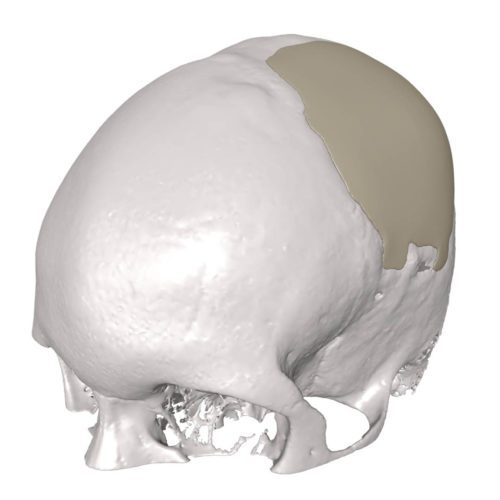
Cranial implant (Image: MedCAD)
These four types of 3D printed patient-specific surgical applications have many benefits: the technology can accurately simulate hard tissues, which is helpful for bone reconstructive surgeries. Additionally, symmetry and functionality are key to successful CMF surgeries, which 3D printing, image processing, and computer software can help virtually model and simulate ahead of time.
“One of the keys for the most benefit for all these types is to understand the concept of mirroring, to find normal tissue to mimic, in order to design the most accurate replica specific to the patient,” the pair wrote. “This involves analyzing the contralateral side that is normal, and using computer software to take its mirror image, thus creating something close to what the ipsilateral anomaly should look like.”
The researchers found that 62% of patients in their review had this mirror technique used in their surgeries.
“Despite the rapid advancement of 3DP in medicine and surgery, 3DP outcomes studies are still in their infancy. Part of this is the lack of a systematic classification of the techniques used, making it more difficult to make comparison studies. Plastic Surgery craniomaxillofacial surgery is an ideal specialty to study patient-specific 3DP surgeries, as the operations are bone-intensive, and 3DP is currently better at simulating hard tissues,” they concluded.
“Our experience so far with patient-specific 3DP for surgery is that it can make surgeries faster, more accurate, and less invasive with smaller scars. This is especially beneficial to pediatric patients where anatomy is finer and more compressed, length of surgery can lead to more blood loss, which is even more critical in children.”
Discuss this and other 3D printing topics at 3DPrintBoard.com or share your thoughts below.
Subscribe to Our Email Newsletter
Stay up-to-date on all the latest news from the 3D printing industry and receive information and offers from third party vendors.
Print Services
Upload your 3D Models and get them printed quickly and efficiently.


You May Also Like
Australia’s AMCRC Funds Titanium 3D Printing R&D
In terms of the global economy’s presently existing state, there is no realistic path to economic resilience that doesn’t start with critical minerals security. This is a problem for pretty...
3DPOD 305: Automating AM with Grenzebach’s Oliver Elbert
Oliver Elbert‘s over ten years in additive manufacturing have been spent automating LPBF. For large, high-volume, or critical parts, Grenzebach has provided custom automation solutions. Depowdering, powder handling, sieving, heat...
AMPulse Asia: Chinese IPOs, Defense Deals, and Dental 3D Printing Lead APAC Roundup
The second half of June brought a wave of additive manufacturing activity across China, Japan, South Korea, India, and Australia. From Chinese IPOs and funding rounds to defense, aerospace, construction,...
Austal, Curtin University and AMCRC Work on R&D Together
Australia’s Additive Manufacturing Cooperative Research Centre (AMCRC) works with 70 industry partners to deliver collaborative R&D projects. They also work on workforce development and technology transfer. It’s kind of analogous...


