Germany: Research Shows Good Response from Students Using 3D Printed Dental Traumatology Training

Authors M. Reymus , C. Fotiadou, R. Hickel, and C. Diegritz explore the uses of 3D printed models in dental traumatology training, with their findings outlined in the recently published ‘3D printed model for hands-on training in dental traumatology.’ For their study, they used an SLA printer to create a 3D printed model of a dental patient’s maxilla, mimicking several different traumatic dental injuries.
Being able to create accurate models exhibiting significant trauma offers a host of benefits to dental students who can take their time in a deliberate learning mode rather than waiting to rush in to see what could be a relatively small number of injured patients on-site. This also accentuates the enormous amount of learning gained from lectures. The hope is that more knowledge can be gained about dental traumatology, as the researchers point out that dental accidents are often treated by general dentists who may not have an adequate education or experience to deal with such cases overall.
The researchers wanted to make a model that was not only realistic but would allow for students to practice both diagnosis and treatment too. They also wanted to design a product that would translate from educational settings to dental clinics. With these hands-on tools available, the authors also created another level to their study regarding the use of dentaltraumaguide.org, offering the resource to only half of the students participating in the study—and comparing their knowledge.
The model was designed and 3D printed as follows to show dental trauma for a 16-year-old boy:
“The data generated were exported as single DICOM files and imported to Invesalius for Mac (Centre for Information Technology Renato Archer, Amarais, Brazil) to convert it into one .stl file. This file was subsequently imported to Meshmixer for Mac 11.0 (Autodesk, San Rafael, CA, USA) and trimmed to a region extending from the right first premolar to the left premolar. The right lateral incisor, the left first incisor as well as the left second incisor were cut out of the STL-mesh and exported as single STL-files.
Using the function ‘Boolean difference’, these teeth were cut out, leaving imitation tooth sockets in their original position. Additionally, the right lateral incisor was positioned at a 30° angle towards the palatal from its original position, and again, the function ‘Boolean difference’ was used to imitate a lateral luxation of the tooth perforating the buccal bone. The left lateral incisor was separated into two parts at its apical third imitating a horizontal root fracture. The extracted left incisor was not changed, imitating an avulsion. The mesial edge of the right incisor was removed, exposing the pulp chamber to imitate a complex crown fracture.”
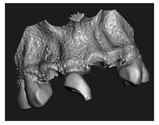
Computer-aided designed model with empty tooth sockets, buccal perforation and complicated crown fracture.
The 32 undergraduate students were tasked to work on the case, even simulating a conversation with the mother of the injured boy as they practiced asking the correct questions about the accident, as well as advising on post-traumatic behavior. Upon examining the 3D printed model, they were given information about every tooth, and asked to offer the following:
- Diagnosis
- Treatment plan
- Recall regime
- Prognosis of each injured tooth
The assessment was considered in these areas:
- Pre-treatment
- Therapy
- Post-treatment
- Recall
- Complications
“The presented workflow allowed the manufacturing of a radiopaque model that imitated a luxation injury, a complicated crown fracture, an avulsion, and a horizontal root fracture in a realistic way,” stated the authors.
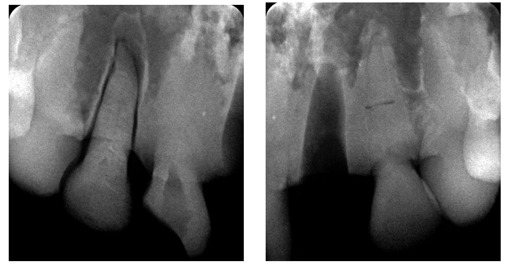
Radiograph of the right lateral incisor with a luxation injury (left) and Radiograph of the empty tooth socket of the left incisor and the left lateral incisor with a horizontal root fracture (right).
And while their goal was for such a workflow to be easily transferred to another dental school, they would need to own a CBCT and a stereolithographic printer, along with software that could be offered free. The 32 students were asked to evaluate the model, with 57 percent reporting it to be ‘very realistic,’ and 43 percent choosing ‘rather realistic.’
“The diagnosis of the lateral luxation was evaluated to be the most difficult of all injuries, whereas the avulsion was the easiest injury to diagnose. Concerning treatment planning, the horizontal root fracture was rated as being the most difficult injury. When listing possible complications, the students had serious problems with the horizontal root fracture.
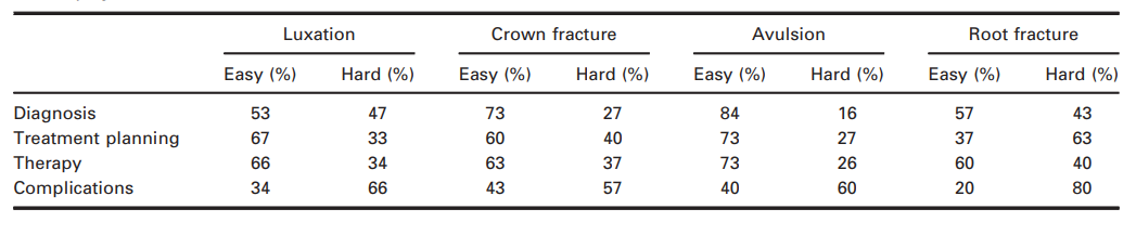
Students’ evaluations of difficulty in diagnosis, treatment planning, therapy and knowledge about complications for each injury.
“All participants reported to have gained new knowledge on dental traumatology, and 97 percent felt better prepared for treating traumatic dental injuries in the future.
“Students seem to focus especially on the diagnosis and treatment of traumatic injuries to teeth when dealing with dental traumatology. This is logical because these steps are of outmost importance for immediate care when confronted with a trauma case. Fortunately, both groups of students in the present study achieved their best results in these fields. The group without access to dentaltaumaguide.org, however, had only poor results when faced with developing a recall regime and knowing about possible complications,” concluded the researchers.
Many dentists and orthodontists rely on 3D printing today for digital dentistry, dentures, and even grafts for issues like alveolar augmentation. What do you think of this news? Let us know your thoughts! Join the discussion of this and other 3D printing topics at 3DPrintBoard.com.
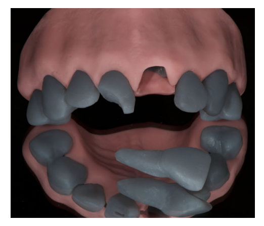
Printed model with gingival mask.
Subscribe to Our Email Newsletter
Stay up-to-date on all the latest news from the 3D printing industry and receive information and offers from third party vendors.
Print Services
Upload your 3D Models and get them printed quickly and efficiently.


You May Also Like
The Stories nScrypt Can’t Tell; and Why That Matters
This article is Part 3 of a three-part series based on 3DPrint.com’s visit to nScrypt’s Orlando headquarters and conversations with Ken Church. There’s an interesting dynamic inside nScrypt’s Orlando headquarters. The...
amsight & toolcraft Improve AM Quality Control for the Semicap Market
As it is in the habit of doing at least once per generation, the semiconductor capital equipment (semicap) market is currently in the process of reinventing itself. This is too...
3D Printing Financials: XTPL Adds New Semiconductor and Defense Customers in Q1 2026
Polish microprinting company XTPL (WSE: XTP) reported first-quarter 2026 revenue of PLN 1.6 million (roughly $441,000) as the company expands into the semiconductor and advanced electronics markets, while also launching...
Creality Marks 12 Years with KliTek and AI-Powered Ecosystem Expansion
For 12 years, Creality has advanced accessible 3D printing technologies, enabling global users to turn ideas into tangible creations. What began as a desktop 3D printer manufacturer has evolved into...