Researchers Create Better Techniques for 3D Printing Surgical Models

 Medical professionals and surgeons around the world have been greatly helped with the emergence and innovation of 3D printed models that not only help pinpoint a diagnosis, but also educate patients and their families regarding health issues (often regarding diseases like cancer and tumors that must be removed). Ultimately, these models can be used by surgeons to practice for delicate surgeries, and then also act as guides in the operating room.
Both US and German scientists have come together now to find improved methods of 3D printing such models and guides, publishing their findings in ‘From Improved Diagnostics to Presurgical Planning: High-Resolution Functionally Graded Multimaterial 3D Printing of Biomedical Tomographic Data Sets.’ While the authors point out how useful anatomical structures are in the medical field, they also explain why 3D printing medical models is currently rife with challenges:
“Typically, features of interest in source biomedical data sets (e.g., crosssectional raster-based biomedical images or volumes from MRI or CT DICOM files) are first isolated from the surrounding tissues, either through a time-consuming manual segmentation approach or through the use of specific intensity thresholds that best capture the feature(s) of interest,” state the authors. “The result of this filtering step is used to generate a single isosurface that is then translated into a printable format [i.e. stereolithography (.stl) file, the current standard for 3D printing workflows], which describes surface geometries through mesh representations of face vertices and normals.”
3D printing of surgical models can be flawed due to reasons like thresholding, or error in digital structure that prevent files from printing and then require users to make additional changes later. The researchers see multimaterial 3D printers as a solution due to their ability to use more than one .stl file:
“This approach greatly expands the number of different anatomical structures that can be represented by different colors or materials (e.g., through the use of multiple thresholds) and, as such, can improve the interpretive value of 3D-printed anatomical models,” state the researchers. “However, the printing of multiple nested .stl files is labor intensive, computationally expensive, and ultimately nonscalable.”
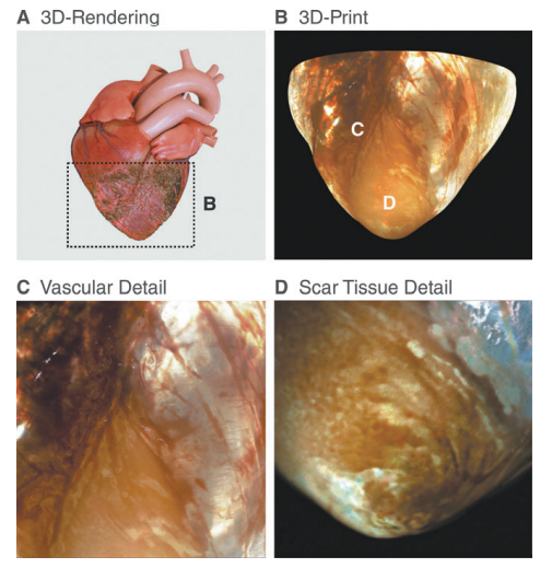
High-spatial resolution 3D-printed structural details in a pathological heart model. (A) Volumetric rendering of an infarcted heart and (B) the corresponding (cropped) 3D print of its apex, clearly showing (C) the structural organization of the superficial coronary vasculature and (D) the details of the left ventricular scar.
As a result, their study is centered around showing how functional bitmap-based workflows are helpful in fabricating biomedical data sets, permitting rapid creation of anatomical models without thresholding. The scientists also ‘bypassed’ more typical 3D printing workflows due to their potential for error, and worked with both MRI and CT data.
[Source / Images: From Improved Diagnostics to Presurgical Planning: High-Resolution Functionally Graded Multimaterial 3D Printing of Biomedical Tomographic Data Sets]“Despite the advantages of this bitmap-based workflow compared with its traditional .stl-based counterpart, there are still some limitations to the technique,” concluded the researchers. “For example, although our 3D-printed models can faithfully replicate the details of their native CT or MRI source files, their ability to successfully depict anatomical features of interest is directly limited by the inherent resolution of each imaging modality.
“Inaccuracies can also be introduced by image reconstruction methods designed to optimize human image viewing rather than 3D printing, and traditional clinical tomography file compression and archival protocols will, in turn, need to be modified as biomedical 3D printing becomes more mainstream. Data set-specific issues also arise with regard to signal intensity and range.”

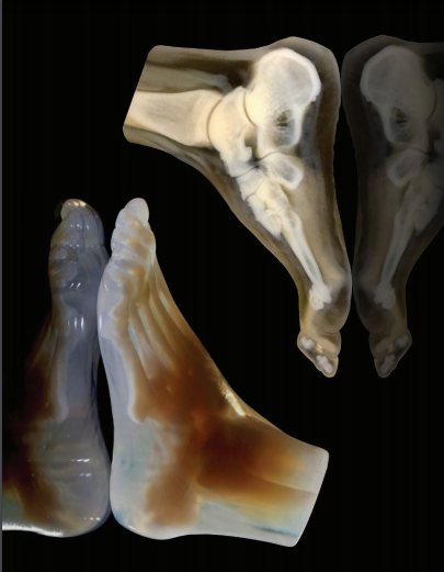
3D printing of scanning electron microscopy (SEM)-based data sets. (A) X-ray transmission image and (B) a corresponding backscattered SEM slice of a healing critical defect in an ovine femur immobilized with a titanium cage implant together with a higher magnification single slice backscattered electron micrograph. (C) A 3D-printed counterpart of the SEM image and (D) the corresponding higher magnification modulus map from the resulting 3D-printed model reveal the subtle differences in mineral density during the early stages of bone healing.
Subscribe to Our Email Newsletter
Stay up-to-date on all the latest news from the 3D printing industry and receive information and offers from third party vendors.
You May Also Like
Printing Money Episode 17: Recent 3D Printing Deals, with Alex Kingsbury
Printing Money is back with Episode 17! Our host, NewCap Partners‘ Danny Piper, is joined by Alex Kingsbury for this episode, so you can prepare yourself for smart coverage laced...
Insights from Cantor Fitzgerald on AM’s Q1 2024 Landscape
A recent survey by Cantor Fitzgerald sheds light on the persistent challenges within the additive manufacturing (AM) industry in the first quarter of 2024. Based on responses from 38 industry...
3D Printing Financials: Xometry’s Scaling up and Strong Start to 2024
Xometry (Nasdaq: XMTR) kicked off 2024 with strong results, boosting its marketplace and technology to new heights. Both revenue and gross margin soared, fueled by an expanding global network of...
3D Printing Financials: Desktop Metal Targets Recovery Amid Net Losses and Revenue Downturn
Despite facing a decline in revenue and the persistent challenges of a tight economic climate, Desktop Metal (NYSE: DM) is making strides toward operational efficiency. The first quarter of 2024...