

Man Helps Save Wife’s Life with 3D Printing – Now Looks to Empower Others with Free ‘3D in Medicine’ Seminar


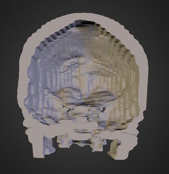
If it were not for forward think surgical team at UPMC and their research in minimally invasive techniques this may not have such a happy ending.Scan of Michael’s wife’s Skull / Tumor
There is nothing scarier in this world, than being told that you or a loved one has been diagnosed with a brain tumor. For one woman, this happened toward the end of last year, but thanks to her husband, Michael Balzer, who was versed quite well in 3D technology, her diagnosis, treatment and recovery went a lot better than expected. For those unfamiliar with Michael Balzer, he is the producer of a podcast and website called, All Things 3D.
“My wife was diagnosed with Menigioma, but the diagnosis was minimized until I was able to review the DICOM information myself and present 3D volume renderings to her to convince her to get another CT with contrast,” Michael Balzer tells 3DPrint.com. “This led to more accurate discovering of [a] 3.5 cm Meningioma in her upper left orbital roof. I was then able to create 3D sagital models, which I then loaded on SketchFab for neurosurgeons to review as we shopped for a neurosurgeon who would remove it using [a] minimally invasive procedure. This process took us three months and we narrowed it down to John’s Hopkins and UPMC (University of Pittsburgh Medical Center).”
Michael then 3D printed two full-scale sagital sections and sent them to the surgeons. UPMC ended up being the only medical center willing to move forward with the new minimally invasive technique, due to the physical models and the imaging that Michael has provided. You can see the 3D scan/model of his wife’s tumor located on the base of her skull below (the ‘after’ model can be seen at the bottom of this article):
In mid-May, an eight hour surgery was performed, through her upper eyelid. Incredibly 95% of the tumor was able to be removed, and just three weeks later, she was back at work, seeing clients again. Michael tells us that she is now almost completely back to normal.
“If it were not for forward think surgical team at UPMC and their research in minimally invasive techniques, this may not have such a happy ending,” said Michael.
Michael still uses 3D scanning technology in order to help provide his wife’s neurosurgeon and optomologist surgeon with periodic updates on her recovery. He does this using a Sturcture Sensor 3D scanner, along with a 4eyes lens attachment product that he sells for the Structures Sensor and iSense scanners. Michael uploaded a 3D model, showing his wife’s progress on Sketchfab, which can be seen below. “Other than the optical distortion, you can clearly see the healing and disparity between eyes, notably the left eye,” Michael explains to us.
“I have not uploaded the latest, but unless you really look now, you would not be able to tell she had any surgery, let alone brain surgery,” explains Michael. “A testament to how far we have come in just five years, where her only recourse would have been for [her] to have 1/3 of her skull removed and brain lifted to remove it. As one neurosurgeon said, this most certainly would [have] caused long lasting problems, including the loss of smell, taste and even the sight in the left eye. 30 years ago she would have died like her mother did at the age of 52. [My wife] just turned 56 eight days ago.”
This goes to show, that perhaps more doctors, surgeons and medical researchers should be using 3D scanning and 3D printing technology, but for one reason or another they are not. That reason is most likely due to the lack of knowledge, and lack of funding for such technology. In Michael’s wife’s case, she was lucky to have a husband as caring and well educated as Michael, complimented by surgeons and a medical center willing to cooperate.
Michael Balzer is grateful for 3D technology, as is his wife. He wants to help spread the knowledge of what 3D technology can do for the medical field, so on October 29th from 6:00 – 8:00 p.m. PST in San Luis Obispo, CA, he will be holding a free seminar titled “3D in Medicine” (site live soon). On hand, will be several 3D medical experts, and the entire presentation will be livecasted via Google Hangouts On Air, via a multiple camera setup. The presentation will show viewers just how medical scanning works, as well as how you yourself can take your own medical scans and turn them into 3D printed models.
More event information can be seen below:
- Location: San Luis Obispo, CA library Main Community Room and via a livecast using Google Hangouts On Air (https://livecast.3dinmedicine.org)
- Date: 10/29/2014
- Time: 6:00 p.m. – 8:00 p.m. Pacific Daylight Time (UTC-7, but I think it goes to PST (UTC-8 the weekend before)
- Contact: info@3dinmedicine.org
- Website: https://3dinmedicine.org
 Preliminary schedule of events:
- 5:30 – 6:30 p.m. – Event Standby and Preparation
- 6:00 – 6:10 p.m. — Introduction of topics and panel of guests
- 6:10 – 6:30 p.m. — Dan Copp, DDS. Overview of how 3D has changed your practice.
- 6:30 – 6:50 p.m. — Scott Klioze, MD. Overview of the latest changes in CT and MRIs that allow for real-time 3D visualization, as well other technologies on the horizon that will change the radiology practice.
- 6:50 – 7:10 p.m. — Jorge Vicente, PhD. Overview of how medical 3D visualization and fabrication tools have changed research and your involvement with CTI and its influence on the world stage.
- 7:10 – 7:30 p.m. — Michael Balzer & Chris Kopack. Tutorial on how import DICOM data into InVesallius, create a volume render and then using the threshold tools quickly create a 3D model that can be imported into 3D modeling app for cleanup and exportation to a 3D printer control program.
- 7:30 – 7:45 p.m. — Chris Kopack. Overview of the creation of the prosthetic for a local boy and the actual prosthetic in use by the boy you created it for.
- 7:45 – 8:00 p.m. — Q/A and Conclusion
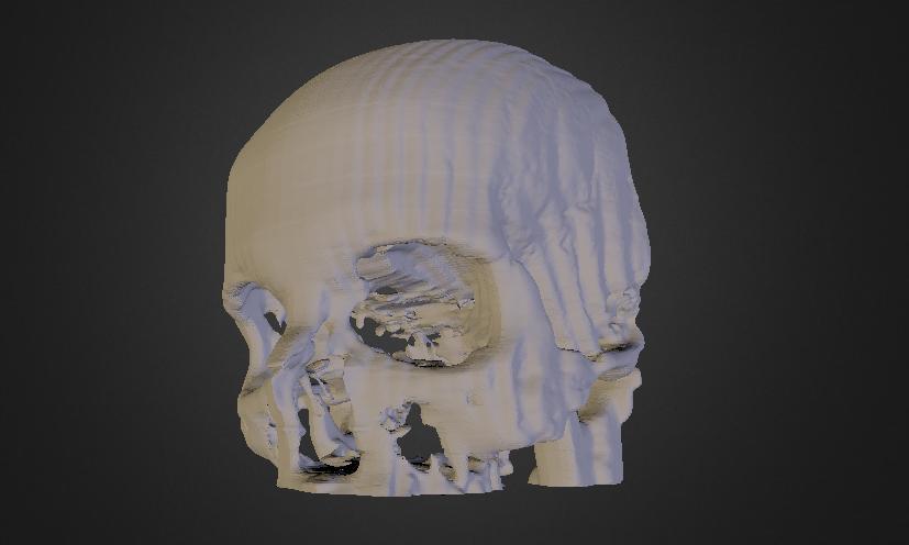
What do you think? Will 3D printing ultimately lead to more successful surgeries? Do you plan on watching or attending this presentation? Discuss in the 3D in Medicine forum thread on 3DPB.com. Check out the post-surgical 3D model of Michael’s wife’s skull below (note the large 3.2cm hole which the surgeon has minimized).
Subscribe to Our Email Newsletter
Stay up-to-date on all the latest news from the 3D printing industry and receive information and offers from third party vendors.
Print Services
Upload your 3D Models and get them printed quickly and efficiently.


You May Also Like
UAS Additive Strategies: Register by June 30 to Learn About the Hottest Topic in 3D Printing
Last week, drone stocks surged on news that the Trump administration is considering a massive investment in the US unmanned aerial vehicle (UAV) industry. Earlier in 2026, the release of...
Stratasys Dental’s Negar Movahed Says They’re “Open for Partnerships”
According to “3D Printing for Dentistry 2025: Market Study and Forecast” by AM Research, the dental 3D printing market generated $5.2 billion in revenue in 2024—that’s nearly one third of...
3D Printing News Briefs, May 30, 2026: RIMPAC 2026, Acquisition, Ceramic Implants, & More
We’re kicking things off with materials news in this weekend’s 3D Printing News Briefs. Then it’s on to a hybrid manufacturing system for a maritime exercise, an expansion of industrial...
3D Printing News Briefs, May 23, 2026: Inserts, Racing, Cultural Heritage, & More
In this weekend’s 3D Printing News Briefs, 3D People has integrated threaded inserts into its online quoting tool, AM Solutions has introduced a more compact solution for automated cleaning and...


