New Research into 3D Printed Bone Implants Could Change the Way We Heal Broken Bones

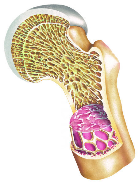
Internal structure of a human bone.
The process of bone formation was discovered back in the 1960s, which led to almost immediate improvements in diagnostic techniques and eventually led to the development of synthetic calcium-based bone repair products. These replacements, called bone grafts, are often made from transplanted bone materials from the patient, cadavers or even some animals. Bone grafts have also been developed using dentin extracted from human teeth that have been ground down into a powder and formed into implants. These days it is far more common for doctors to use a material that will eventually break down in the body and be replaced by the newly grown bone tissue, although metal implants are occasionally required with serious breakage.
For years, bone grafts have been a common method of helping patients with broken or damaged bones to properly heal. Because bone tissue can quickly heal itself, an implant, or scaffold, is used to surgically hold all of the damaged bone fragments together and act as a bridge of sorts that will ensure that they heal correctly. Recently doctors have started 3D printing these implants in order to ensure that the scaffold is sized and fit exactly to the patient’s specific needs. However doctors are limited in the materials that they can use to 3D print bone grafts due to the need for the implants to be strong enough to offer structural support.
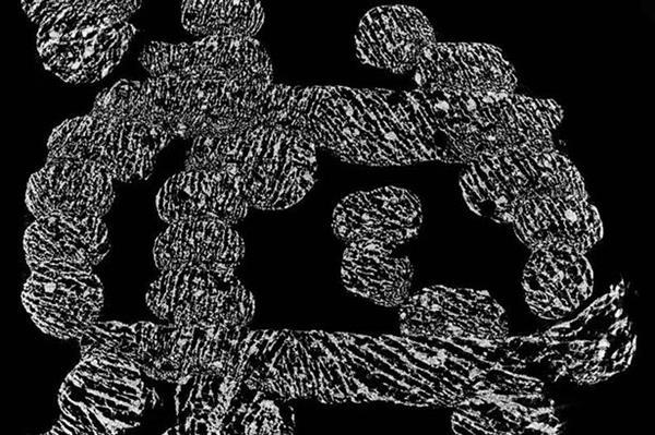
A micro-computed tomography image of the 3D printed bone scaffold. [Image: NTU]
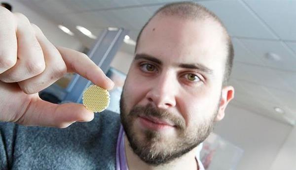
Nottingham Trent University researcher Manolis Papastavrou with a 3D printed bone scaffold. [Photo: NTU]
“This research demonstrates how 3D printing in combination with freezing can reduce significantly the fabrication time and cost of such medical devices. The secret behind the toughness of many biological materials is the way their components are arranged from the molecular all the way up to a macro level. Using this design strategy could help engineer bone scaffolds, whose porosity does not compromise their strength. In the long term, this research could contribute in replacing the use of metal in orthopaedic implants with materials that can be broken down by the body,” explained PhD candidate and researcher Manolis Papastavrou.
![]() The exact materials used to create 3D printed bone grafts vary from manufacturer to manufacturer, however they all contain many of the same materials found in natural bone. They typically are made from ceramics with high levels of calcium phosphates, but also can be made using bioglass or calcium sulphate. All of these materials are biologically active and will eventually be dissolved or absorbed into the patient’s body. While the materials would naturally dissolve as new bone tissue grew, if the scaffold was too thick or dense it could prevent the bone from healing correctly.
The exact materials used to create 3D printed bone grafts vary from manufacturer to manufacturer, however they all contain many of the same materials found in natural bone. They typically are made from ceramics with high levels of calcium phosphates, but also can be made using bioglass or calcium sulphate. All of these materials are biologically active and will eventually be dissolved or absorbed into the patient’s body. While the materials would naturally dissolve as new bone tissue grew, if the scaffold was too thick or dense it could prevent the bone from healing correctly.
Until this new breakthrough, due to the need for the implants to be porous enough to encourage new bone growth, the strength of the scaffolding was an ongoing issue. The new 3D printed bone microstructures combined with the freezing process developed by Papastavrou and the rest of the researchers at Nottingham Trent’s Design for Health and Wellbeing Research Group was recently presented at a conference titled Printing for the Future. The day long presentation event took place on January 19th 2016 at the Institute of Physics, London. Discuss your thoughts on these new materials in the 3D Printed Implants With Crystals forum over at 3DPB.com.
Subscribe to Our Email Newsletter
Stay up-to-date on all the latest news from the 3D printing industry and receive information and offers from third party vendors.
Print Services
Upload your 3D Models and get them printed quickly and efficiently.


You May Also Like
Roboze Opens U.S. Aerospace & Defense Headquarters in El Segundo
The manufacturing sector is made up of clusters: “geographic concentrations of interconnected companies” that both cooperate and compete with each other. Of course, this is true about any sector in...
EOS Invests $3M In Its Texas Manufacturing & Logistics Facilities to Serve North American Customers
The trajectory of reshoring under President Trump has been largely a mixed bag so far. While tariffs still seem to be doing more harm than good to the U.S. domestic...
At AIAA SciTech 2026, 3D Printing Was Part of the Workflow — Part I
The AIAA SciTech Forum 2026 brought much of the aerospace community together in one place. With roughly 6,000 attendees, 115 exhibitors, 21 sponsors, and nearly 3,000 technical paper presentations, the...
€73 Million Investment Round into SWISSto12
SWISSto12 is on the move. I think that the firm owns one of the most promising and profitable applications for 3D printing, RF components, and it is showing real growth...