Spectrum Health 3D Prints the First Heart Model Using Multiple Imaging Techniques

![]() Surgeons have been using 3D printed models of their patients’ hearts for preoperative planning and strategizing for a while now. While not yet quite routine, the practice has still become a relatively common process for difficult, complex or excessively dangerous heart surgeries. The models printed for these procedures can often give doctors greater insight into what they will see when they open a patient up, and dramatically reduce the length of the operation by eliminating the need for surgical probing or exploring the site of the defect being repaired. However, the 3D model is only as good as the data used to create it, and currently no singular method for obtaining a 3D model of a heart is perfect.
Surgeons have been using 3D printed models of their patients’ hearts for preoperative planning and strategizing for a while now. While not yet quite routine, the practice has still become a relatively common process for difficult, complex or excessively dangerous heart surgeries. The models printed for these procedures can often give doctors greater insight into what they will see when they open a patient up, and dramatically reduce the length of the operation by eliminating the need for surgical probing or exploring the site of the defect being repaired. However, the 3D model is only as good as the data used to create it, and currently no singular method for obtaining a 3D model of a heart is perfect.
But heart specialists at Spectrum Health, a group of 12 not-for-profit hospitals in West Michigan and considered one of the best healthcare providers in the country, has just 3D printed the first model of a patient’s heart using combined data from imaging techniques. The hybrid 3D printed model has significantly more detail than models created using standard techniques. Spectrum Health is the first healthcare provider to develop a process to combine virtual data from multiple imaging scans.
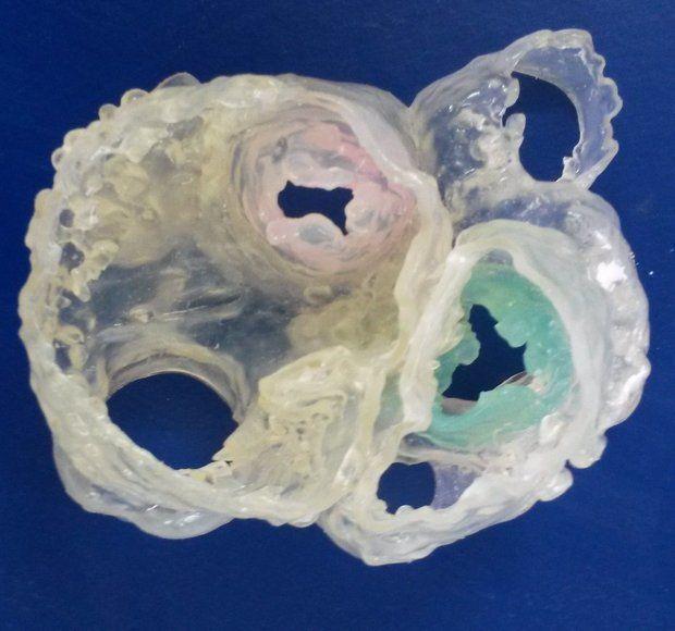
3D heart model created by combining CT scan data and 3D transesophageal echocardiography data
Typically when a surgeon needs a 3D model of a patient’s heart they use one of several methods available to them. In the last year we have seen hearts 3D printed using data from CT scans, MRI data and recently even an ultrasound process called 3D echocardiography. This 3D model printed by Spectrum Health combined the data gathered from a CT scan and using 3D echocardiography. Spectrum Health is also further developing the new process to incorporate highly detailed data from an MRI scan.
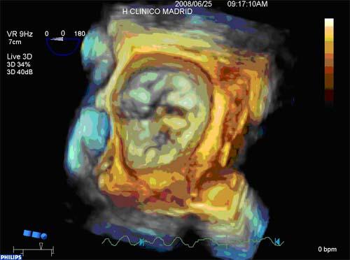
Mitral valve prolapse scan collected using 3DTEE imaging
“Hybrid 3D printing integrates the best aspects of two or more imaging modalities, which can potentially enhance diagnosis, as well as interventional and surgical planning. Previous methods of 3D printing utilize only one imaging modality, which may not be as accurate as merging two or more datasets,” cardiac sonographer and the lead author of the proof-of-concept study Jordan Gosnell explained to Mlive.com.
Each of the various imaging techniques used in this hybrid process has different strengths, and by combining data from multiple techniques doctors can create an extremely detailed 3D model of the patient’s heart. The CT scan is used to collect highly detailed data of the exterior anatomy of the heart. The inside of the heart is visualised using an MRI, which can also generate detailed scans of its musculature. And finally the 3D transesophageal echocardiography (3DTEE) process focuses specifically on the heart valves, the primary location of the type of defects that the process was developed to correct.

Dr. Joseph Vettukattil, interventional cardiologist with Spectrum Health’s Congenital Heart Center
The final proof-of-concept study for this process is being presented this week at CSI 2015 (The Catheter Interventions in Congenital, Structural and Valvular Heart Disease Convention) in Frankfurt, Germany. The presentation will be handled by the study’s senior author, Dr. Joseph Vettukattil, one of the leading heart specialists in the world. He has also helped develop the process of generating 3D models using 3DTEE, specifically for use with patients diagnosed with congenital heart disease. While the new process has already proven to be effective, Dr. Vettukattil does say that they still need to conduct additional research before determining the role of these hybrid 3D models in making surgical decisions. You can discuss this new medical advancement over in our 3D Printed Heart Model Using Multiple Imaging Techniques forum thread at 3DPB.com.
Subscribe to Our Email Newsletter
Stay up-to-date on all the latest news from the 3D printing industry and receive information and offers from third party vendors.
Print Services
Upload your 3D Models and get them printed quickly and efficiently.


You May Also Like
Harvard SEAS Engineers Develop 3D Printing Method for Soft Robotic Components with Programmable Shapes
The world of soft robotics is still largely in its pure research phase, but the R&D landscape has started to produce examples of early-stage commercialization. Researchers have started to refine...
3D Printing News Briefs, February 14, 2026: Project Call, Maritime Construction, Prosthetics, & More
Happy Valentine’s Day! We’re starting this weekend’s News Briefs off with a Project Call award, and then moving on to a business growth program. We’ll end with research in underwater...
CASF: A Green Surface Finishing Technology for AM Hard Metal Alloys and Fatigue Improvement
Sugino Machine Ltd has recently completed development of a highly specialized surface-finishing technology capable of removing partially melted particles, debris, and alpha case left behind by additive-manufactured (AM) laser powder...
Hardware is Dead. Here’s What Actually Wins in Additive Manufacturing.
Hardware is rapidly commoditizing across additive manufacturing. Specifications have converged. Price competition has intensified. Margins have compressed. For companies attempting to scale additive manufacturing beyond prototyping, this shift has profound...