
Patient-Specific Anatomical Models for 3D Printing and AR/VR


Researchers from the US and Canada have released a supplement to their Radiological Society of North America (RSNA) 2018 hands-on 3D printing course, reviewing techniques for creating 3D printed cranio-maxillofacial (CMF), orthopedic, and renal cancer models. These models can also be viewed in both virtual and augmented realities, as outlined in their recently published ‘Creating patient-specific anatomical models for 3D printing and AR/VR: a supplement for the 2018 Radiological Society of North America (RSNA) hands-on course.’
As 3D printing becomes more accessible, affordable, and less intimidating to users in the mainstream, hospitals and doctor’s offices are beginning to use this technology on-site and on-demand in offering more patient-specific care—whether in the creation of models or medical devices. Both augmented reality (AR) and virtual reality (VR) tend to accompany 3D design and printing also, especially in the medical field where visualization is key in making diagnoses, planning treatment, and educating patients, their families, and medical students too.
“In order to efficiently create 3D printed anatomic models and to use them safely for medical purposes, radiologists and medical professionals must understand the process of converting medical imaging data to digital files,” state the authors.
“Therefore, to educate radiologists and other medical professionals about the steps required to prepare DICOM data for 3D printing, hands-on courses have been taught at the Radiological Society of North America (RSNA) annual meeting since 2014.”
Their courses are for educational purposes only, and not the promotion of any products, emphasizing the use of ‘FDA-cleared software,’ with examples in this case representing craniomaxillofacial (CMF), orthopedic, and renal cases.
In creating optimized models, suitable parameters must be considered, along with the following:
- Spatial resolution (approximately 1 mm3)
- Reconstruction kernel
- Multi-phase contrast
- Metal artifact reduction
- Sequence parameters for MRIs
As a note, it is generally not cost-effective to perform repeat imaging—along with presenting concerns regarding excess exposure to radiation.

Stages of the anatomical modeling process
Image processing was provided by Mimics inPrint, allowing the researchers to fabricate anatomic regions of interest from the DICOM data. Workflow was comprised of the following steps:
- Create ROI
- Edit ROI
- Add part
- Edit part
- Prepare print
The main tools used include:
- Zoom
- Pan
- Scroll
- Zoom
- Navigate with one-click
- Threshold adjustment
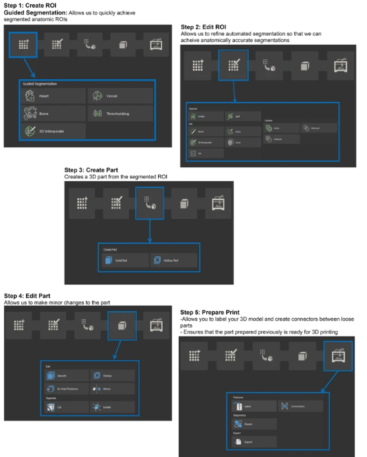
Mimics InPrint workflow steps including 1) Create ROI, 2) Edit ROI, 3) Add Part, 4) Edit Part, and 5) Prepare Print
Three cases were followed:
Pelvic fracture – with the 3D printed model, the researchers were able to help surgeons visualize features of the bones fragments and strategize regarding any impending surgery.
“3D printed pelvic models can also lead to improved perioperative outcomes as compared to patients treated with conventional pre-operative preparation,” stated the researchers. “Mirror images of the opposite intact hemi-pelvis may also be created and can be used to pre-contour fixation plates and these have been reported to reduce surgical times.”
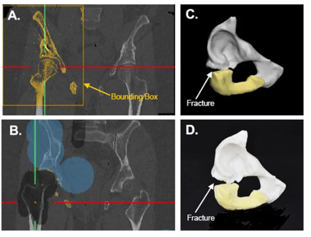
a Coronal CT image showing threhsolded right pelvic bones, showing similar colors for the pubis, ischium, and femur. b Coronal CT image showing splitting of the pelvis (blue) from the femur (black). c 3D computer model showing the pubis (white) and ischium (yellow). d Photograph of 3D printed model
Mandible tumor – 3D printed models help everyone involved understand the anatomy of the mandible, from the patient to the doctor to the medical student and may also serve as surgical pre-planning guides.
“To best depict this anatomy, we chose to print using material jetting (Stratasys J750, Eden Prairie, MN) with the mandible transparent and the tumor and nerves in high presence colors such as blue and green,” stated the researchers. ‘The total print time for this model was 9 h and 24 min using a high mix print setting.”
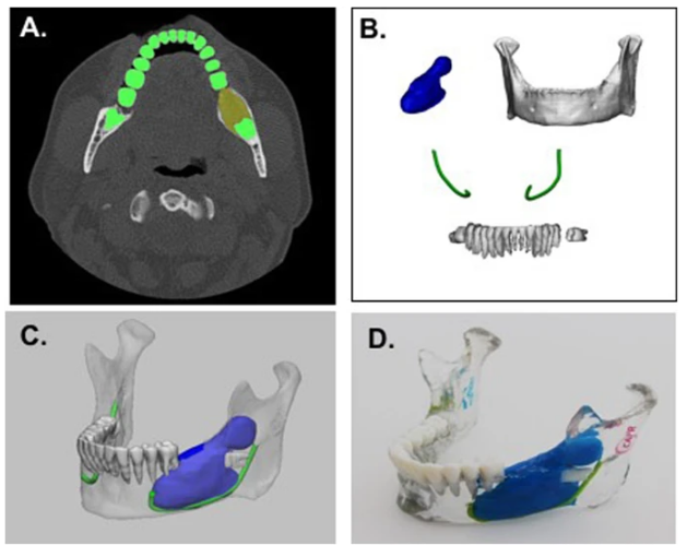
a Axial CT image showing segmentation of teeth (green) and tumor (yellow). b 3D anatomical regions of interest including the tumor (blue), mandible (white), teeth (white), and nerves (green). c 3D visualization of model including all anatomical parts. d 3D printed mandible tumor model including the mandible (clear), teeth (white), tumor (blue), and nerves (green)
Kidney tumor – 3D printed models can be used for both the nephrectomy process as well as planning ablative therapy. The model can be used for evaluating the status of the tumor, as well as its proximity to other anatomy. The model can be used for training purposes as well as helping reduce time in the operating room.
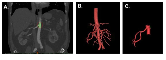
a Coronal CT image showing aorta and right renal artery selection. b 3D visualization of segmented arterial structures. c Remaining arterial region after trimming has been performed
“AR or VR models can be created for all the cases list too, exported and then prepared in Unity,” concluded the researchers.
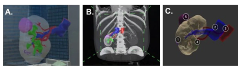
3D kidney tumor model visualized a in AR using the HoloLens AR headset (Microsoft, Redmond, WA), b in VR using Syglass software (Syglass, IstoVisio, Inc., Morgantown, WV) in combination with the Oculus Rift (Facebook, Menlo Park, CA), and c in VR using the Sketchfab app (Sketchfab, New York, NY) and a smartphone device. Each structure is numbered so that the unfamiliar user can easily identify each individual structure: 1 – kidney, 2 –vein, 3 – artery, 4 – collecting system, 5 – renal tumor
3D printed medical models have added not only a huge educational quotient in terms of teaching patients, medical students, and more, but also allow surgeons to pinpoint tumors, plan for hip surgeries, healing fractures, educating aneurysm patients, and more.
What do you think of this news? Let us know your thoughts! Join the discussion of this and other 3D printing topics at 3DPrintBoard.com.
[Source / Images: ‘Creating patient-specific anatomical models for 3D printing and AR/VR: a supplement for the 2018 Radiological Society of North America (RSNA) hands-on course’]Subscribe to Our Email Newsletter
Stay up-to-date on all the latest news from the 3D printing industry and receive information and offers from third party vendors.
Print Services
Upload your 3D Models and get them printed quickly and efficiently.


You May Also Like
Excellent Desktop Injection Molding, Made in Italy by Robot Factory
I was captivated when I saw my first Robot Factory 3D printer. The robust, precise machine was built to last. And this was in an era of very flimsy, disposable,...
Pogačar & Fairlight Cycles Show Us Low Cost 3D Printed Components for Bikes
There has been a lot going on in 3D printing for bicycles over the years. The most successful implementation so far is in bicycle seats. Carbon 3D printed seats are...
3D Printing News Briefs, June 18, 2026: Reseller, Relocation, Metal Space Powder, & More
We’ll start with business news in today’s 3D Printing News Briefs, as XJet appointed a value-added reseller in Germany, BIO INX is expanding its presence in the Italian market, and...
Researchers Combine AI and Bioprinting to Create Tiny Blood Vessel Networks
If 2026 has a theme in bioprinting, it may be blood vessels. Researchers can already print incredibly sophisticated tissues. The harder part is keeping those tissues alive. Without a network...


