Miniature to Massive: 3D Printed Cells, Viruses and Parasites Demystify Microbiology


One of the most terrifying things about a deadly virus like Ebola, particularly potent influenza strains or, say, the Hantavirus has been the fact that, while they can potentially kill millions of people we cannot actually even see them. It is like fighting an invisible enemy, a prospect that makes such a battle even more frightening and the foe more formidable.
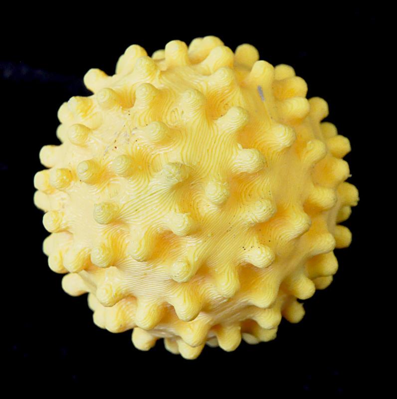
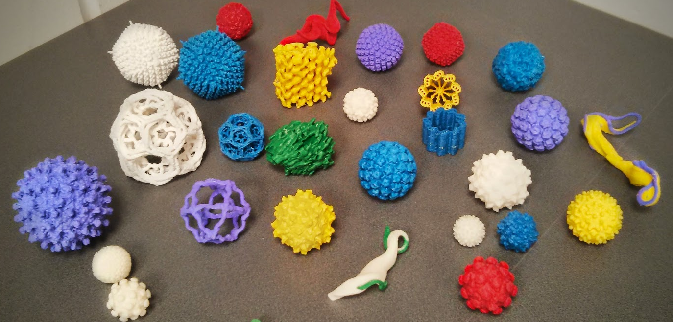
3D-printed, larger-than-life model of a Hepatitis B cell.
Recently, we reported on a couple of makers who, whether in a tongue-in-cheek gesture or otherwise, created 3D printed, larger-than-life images of the Ebola virus. However, in a clearly educational effort, the Bioimaging Unit at Oxford Brookes University has gone much further, presenting an exhibition titled “Miniature to Massive: The Micro-World in Your Hands.” The exhibition, held at London’s Museum of Natural History from November 14 to 16, highlighted some of the work done in the Bioimaging Unit at Oxford Brookes. The exhibition was part of the larger Biotechnology and Biological Sciences Research Council’s Great British Science Festival, a prestigious event where scientific teams showcase their work.
The exhibition featured 3D models of organisms like viruses and parasites. Visitors could view organisms through microscopes, look at 3D photographs of a variety of organisms using 3D glasses, and inspect 3D printed models that are many times larger than life. One member of the Bioimaging Unit exclaimed, “…The first day of the exhibition was a schools day and 1,500 local schoolchildren came around the whole tent and had an excellent day looking at our 3D printers, feeling the models, getting a feel of what’s  going on inside cells, and generally getting engaged with biology.”
The Oxford Brookes Bioimaging Unit uses 3D microscopy to study the interior workings of cells. Using advanced microscopes from which they can 3D print organisms has been a groundbreaking effort headed by Dr. Louise Hughes, who oversees the Unit and has recently been awarded a New England Bioscience, Passion in Science prize. The prestigious award salutes Dr. Hughes’ important work in exploiting the possibilities of 3D printing not only in the lab setting, but to bring microscopy to the general public, including the visually impaired.
A video of the Miniature to Massive exhibition shows children handling brightly-colored 3D models of gigantic (relatively speaking) organisms — and having fun. “It’s a fantastic opportunity,” said Dr. Hughes, “[for the public] to come and see what we scientists do, to see the passion we have.”
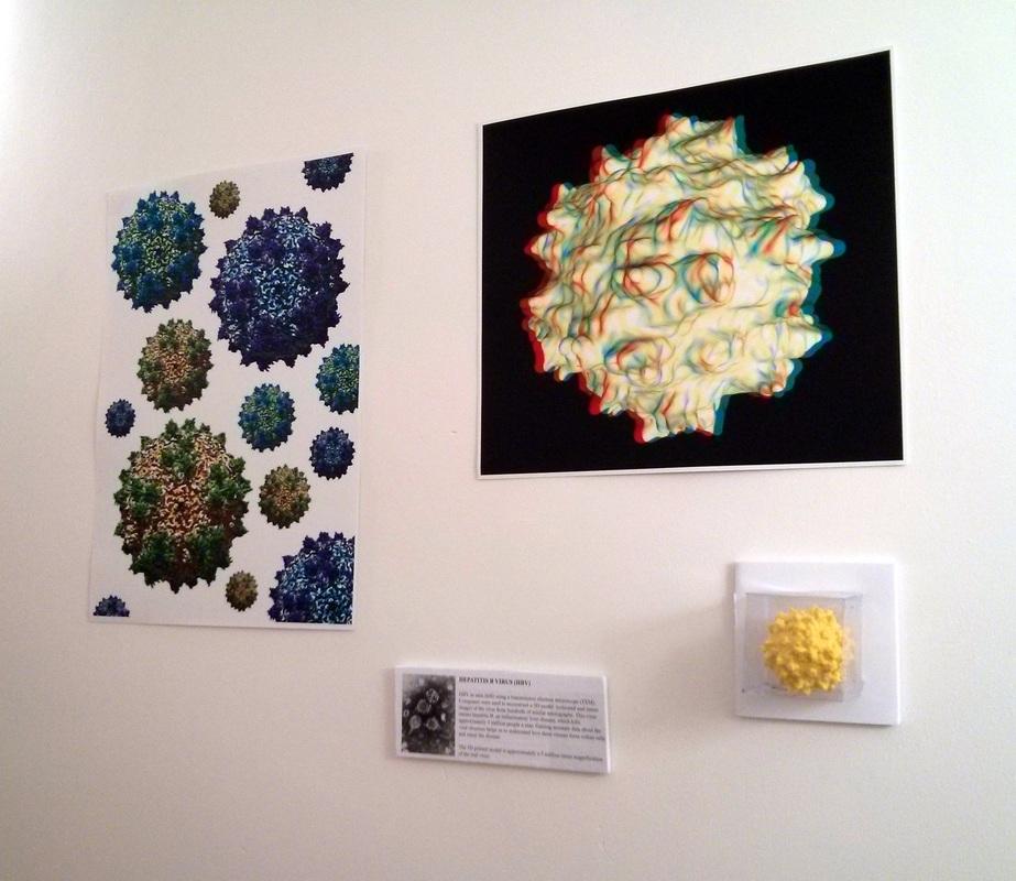
The exhibition included displays of 3D microimaging; visitors could don 3D glasses and get the full picture.
Dr. Hughes also explained how the use of 3D models has enhanced the scientific research process, describing situations where having a 3D printed model has facilitated a more extensive exploration of an organism and the interpretation of related data. Researchers have in some instances come up with new approaches to studying certain organisms based on their altered perceptions of an organism’s structure after seeing an enlarged 3D model.
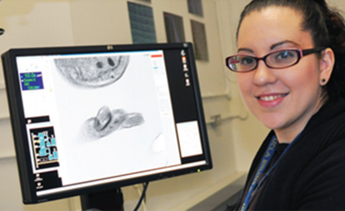
Dr. Louise Hughes
Whether children or savvy scientific researchers, it seems that 3D imaging and printing has the power to demystify the microscopic, to enable us to visualize what was previously invisible to the naked eye and thus more frightening. Getting face-to-face with the microscopic world around us and inside of us is one of the remarkable achievements of 3D technology, when we can actually touch microscopic data via 3D models of viruses, cells, and parasites. It’s a brave new 3D printed world!
Would you go to an exhibit like this one? Let us know what you think of the benefits of seeing viruses made visible in the Miniature to Massive forum thread over at 3DPB.com.
Subscribe to Our Email Newsletter
Stay up-to-date on all the latest news from the 3D printing industry and receive information and offers from third party vendors.
Print Services
Upload your 3D Models and get them printed quickly and efficiently.











You May Also Like
AM Investment Strategies 2026 Sets the Stage for AMS 2027
AM Investment Strategies 2026 Nov. 5 · 12 p.m. ET · Free online event RESERVE YOUR SPOT ↗ A one-hour online discussion featuring some of the biggest names in 3D...
With Its Latest $200M Round, ICON May Be Running Away With the 3D Printed Construction Market
I used to view ICON, the Austin-based additive construction (AC) firm, as one of a handful of companies that could realistically consider itself the leader of the 3D printed construction...
How Should Businesses Build New Business? Dirk Simon Weighs In — Part 1
Dirk Simon had deep experience in materials and chemistry before joining the 3D printing industry. Instrumental in BASF and Farsoon’s market introduction, Dirk has spent a lot of time thinking...
3D Printing Services Near $2.5B as AM Market Shifts Beyond Hardware
The additive manufacturing market may be growing again, but one of the more interesting numbers behind that recovery has little to do with selling 3D printers. AM services generated nearly...