
Bioprinting 101 – Part 13, Imaging Technology


We talked about the way phantoms are created within bioprinting in our previous article. In this article, we will focus more on imaging technology that makes all of this possible. The medical field has been using imaging technology for quite some time for diagnosis, prognosis, as well as general healthcare applications. The scope of imaging technology has been widened with the interaction it is having with bioprinting, and we will look into imaging processes and how they affect this field.
Why is imaging so important for bioprinting you may ask? There is a large amount of data that is processed within an image. Images have resolution values that are determined by pixelation data. In order to process an image, one must digitize the image into a pixelated form. Pixelated images are the key to 3D models. In order to have a precise 3D model of a heart, we must have very high-resolution images within the 2D sphere. 2D images are stitched together to create a 3D transformed object. In terms of imaging technology, the better the original 2D images, a 3D object stitched from said images will have a higher quality in the overall look. When it comes to a 3D bioprinted object or phantom, we want to make sure that it has high fidelity and 3D image quality, thus the precision of different imaging technologies becomes very critical. We will talk about the advantages and disadvantages of each imaging technology in terms of bioprinting.
A computerized tomography (CT) scan combines a series of X-ray images taken from different angles around your body and uses computer processing to create cross-sectional images (slices) of the bones, blood vessels and soft tissues inside your body. CT scan images provide more-detailed information than plain X-rays do. A CT scan has many uses, but it’s particularly well-suited to quickly examine people who may have internal injuries from car accidents or other types of trauma. A CT scan can be used to visualize nearly all parts of the body and is used to diagnose disease or injury as well as to plan medical, surgical or radiation treatment. To bioprint with a layer-by-layer approach, tomographic reconstruction is done on the images. Tomographic reconstruction is a type of multidimensional inverse problem where the challenge is to yield an estimate of a specific system from a finite number of projections. The mathematical basis for tomographic imaging was laid down by Johann Radon. The system we are dealing with is the 3D dimensional object that is created through rotational geometric analysis on the multiple 2D images we have taken with a CT scanner. The now-2D images are then sent to the printer to be made. These cells are then mixed with a special liquefied material that provides oxygen and other nutrients to keep them alive. In some processes, the cells are encapsulated in cellular spheroids 500μm in diameter. This aggregation of cells does not require a scaffold, and are required for placing in the tubular-like tissue fusion for processes such as extrusion.
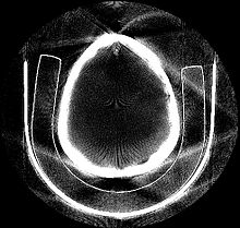 Tomographically Reconstructed Image
Magnetic resonance imaging (MRI) is a medical imaging technique used in radiology to form pictures of the anatomy and the physiological processes of the body in both health and disease. MRI scanners use strong magnetic fields, magnetic field gradients, and radio waves to generate images of the organs in the body. MRI does not involve X-rays or the use of ionizing radiation, which distinguishes it from CT or CAT scans and PET scans. Magnetic resonance imaging is a medical application of nuclear magnetic resonance (NMR). NMR can also be used for imaging in other NMR applications such as NMR spectroscopy.
Diagnostic ultrasound, also called sonography or diagnostic medical sonography, is an imaging method that uses high-frequency sound waves to produce images of structures within your body. The images can provide valuable information for diagnosing and treating a variety of diseases and conditions. Most ultrasound examinations are done using an ultrasound device outside your body, though some involve placing a device inside your body. Diagnostic ultrasound is a safe procedure that uses low-power sound waves. There are no known risks. Ultrasound is a valuable tool, but it has limitations. Sound doesn’t travel well through air or bone, so ultrasound isn’t effective at imaging body parts that have gas in them or are hidden by bone, such as the lungs or head. To view these areas, your doctor may order other imaging tests, such as CT or MRI scans or X-rays.
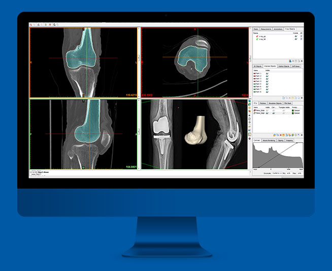
We need to explain specific software that helps to stitch images into 3D files. That software is called Mimics. Materialise Mimics is an image processing software for 3D design and modeling, developed by Materialise NV, a Belgian company specialized in additive manufacturing software and technology for medical, dental and additive manufacturing industries. Materialise Mimics is used to create 3D surface models from stacks of 2D image data. These 3D models can then be used for a variety of engineering applications. Mimics is an acronym for Materialise Interactive Medical Image Control System. It is developed in an ISO environment with CE and FDA 510k premarket clearance.
A major development within bioprinting is the development of imaging technology. Radiology and imaging allows for precision image quality that leads to overall higher resolution 3D models of structures of the body. At the previous RSNA conference in 2018, various 3D printing companies were in attendance showing the importance of Radiology and imaging technology when it comes to the future of 3D printing. This shows the way the industry is trending, and it will be interesting to see what can occur within the future. Precision is very important to the future of bioprinting. It will be cool to see how exact we can become with imaging and 3D model reconstruction.
Subscribe to Our Email Newsletter
Stay up-to-date on all the latest news from the 3D printing industry and receive information and offers from third party vendors.
Print Services
Upload your 3D Models and get them printed quickly and efficiently.


You May Also Like
3DPOD 302: Digital Inventory for AM with Mikhail Gladkikh, Würth Additive Group
Mikhail Gladkikh has worked in oil and gas for many years. With this background, we obviously talk about energy market turbulence and the adoption of AM in oil and gas....
Spectrum Filaments Gets Investment: How They Could Win in Filament
Spectrum Filaments is a long-time high-quality filament supplier based in Poland. With good tolerances, roundness, and consistency coupled with affordable pricing, the firm has been a mainstay for makers, industrial...
NX Atomics and Sciaky Collaborating to 3D Print Nuclear Components
For decades, the nuclear industry has quietly experimented with and implemented additive. Bouyed by the likes of ORNL, companies such as Westinghouse have 3D printed components serially. We have an...
Incodema3D Buys 14 Metal EOS Systems, Now One of the World’s Largest Metal 3D Printer Operators
Recently, a majority stake of 3D printing service bureau Incodema3D was purchased by AFM Capital. Under new ownership, the Freeville, New York company is now using its cash-rich parent for...


