Researchers Compare Human Cadavers and 3D Printed Anatomical Models to Determine Print Accuracy


3D pelvis model in Meshlab.
Surgeons often turn to innovative technology, like 3D printed anatomical models, to get a closer, more detailed look inside a patient’s body ahead of complex procedures. In particular, these models can help trauma surgeons determine the best approach to fixing complex fractures. But just how accurate are these 3D printed models when it comes to matching the look of human bone?
Accuracy is very important when it comes to the fitting of surgical guides and plates, as it’s difficult to characterize and analyze these fractures ahead of time, even with the help of CT scans. But a collaborative group of researchers from the Netherlands just completed a validation study to test the accuracy of 3D printed anatomical models for surgical planning purposes.
Their results were published in a paper, titled “Validation study of 3D-printed anatomical models using 2 PLA printers for preoperative planning in trauma surgery, a human cadaver study,” in the European Journal of Trauma and Emergency Surgery; co-authors are Lars Brouwers from Elisabeth-Tweesteden Hospital, Arno Teutelink with Bernhoven Hospital, and Fiek A. J. B. van Tilborg, Mariska A. C. de Jongh, Koen W. W. Lansink, and Mike Bemelman from Elisabeth-Tweesteden Hospital.
“Surgeons generally need years of practice to transform a two-dimensional (2D) image into a three-dimensional (3D) image in their mind in order to get a proper understanding of the fracture patterns. CT software however easily enables volume rendering of 2DCT into a 3D reconstruction,” the study’s introduction reads.
“3D printing has become increasingly utilized in the preoperative planning of clinical orthopaedics, trauma orthopaedics and other disciplines over the past decade [2]. 3D-printed models are readily accessible due to the wide availability of 3D printing techniques and 3D printers. 3D printing contributes to a better understanding of the surgical approach, reduction and fixation of fractures, especially in complex fractures such as acetabular fractures.
“However, it is unclear how a 3D-printed model relates to a human bone. To our knowledge, there is no literature that validates the accuracy of 3D-printed models in a preoperative planning strategy when applied to real human bones.”
-
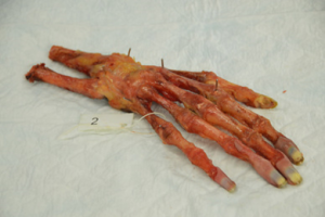
- Cadaver hand with marker points.

-
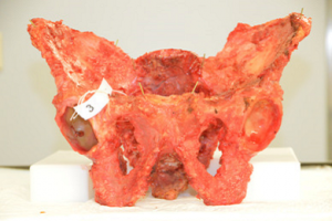
- Cadaver pelvis with marker points.

-
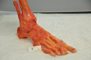
- Cadaver foot with marker points.
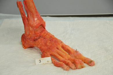
The team dissected nine human cadavers to acquire three specimens each of a pelvis, hand, and foot, and inserted Titanium Kirschner (K-) wires in them to mark important anatomical landmarks. In order to convert CT scans in the DICOM file format to STL, the team used a Siemens Somatom Definition AS 64-slice CT to scan the specimens at a slice thickness of 0.6 mm, before moving on to the next stage of image post-processing.

3D model of a pelvis after CT scanning with all measurements between the five marker points performed on the Philips Intellispace Portal.
Phillips Intellispace Portal software was used to render the the DICOM data into 3D reconstructions, and then the data was digitally cleaned and saved as STL files, the landmarks of which were measured by two independent reviewers using open source Meshlab. The files were then imported and the G-code generated, and then the models were 3D printed, in a ratio of 1:1, on both an Ultimaker 3 and a Makerbot Replicator Z18 using PLA material.
Then, the independent observers measured the distances between the K-wires on the 3D printed models and the human cadaver specimens, in addition to Meshlab, 2DCT, and the 3D reconstructions. These distances were measured a second time one month later, with the exception of the specimens, as these had to be disposed of quickly. In addition to analyzing the observers’ data, the team also completed some calculations to provide an overview of the print process settings.
According to the study, “The least decrease in average distance in millimetres was seen in “the 3D printed pelvis 1”, − 0.3 and − 0.8% on respectively the Ultimaker and Makerbot when compared with cadaver Pelvis (1) The 3D model of “Hand 2” showed the most decrease, − 2.5 and − 3.2% on the Ultimaker and Makerbot when compared with cadaver hand (2) Most significant differences in measurements were found in the conversion from 3D file into a 3D print and between the cadaver and 3D-printed model from the Makerbot.”

Cadaver hand with titanium K-wire marker points next to its 3D printed model. The K-wires are visible on the 3D printed model.
The team concluded that 3D printing can be used to create accurate medical models that are “suitable” for pre-op planning; they also determined that the Ultimaker 3 was just a little more accurate than the Replicator Z18. The researchers recommend that any medical professionals who use 3D printed models for surgical planning first test out the accuracy of their own 3D printing processes.
Discuss this and other medical 3D printing topics at 3DPrintBoard.com, or share your thoughts below.
[Images: Brouwers et. al.]Subscribe to Our Email Newsletter
Stay up-to-date on all the latest news from the 3D printing industry and receive information and offers from third party vendors.
Print Services
Upload your 3D Models and get them printed quickly and efficiently.


You May Also Like
3D Printing News Briefs, June 4, 2026: Anniversary, Maritime, Marine Organisms, & More
In today’s 3D Printing News Briefs, Snapmaker is celebrating 10 years with a series of updates. XJet is collaborating with eqops for sales and support across the UK and Ireland,...
3D Printing News Briefs, April 22, 2026: DINOs, Post-Processing, AM for Aerostructures, & More
We’ll start with event news in today’s 3D Printing News Briefs, as AMUG presented its DINO Award to six members at this year’s conference, and Axtra3D celebrated its five-year anniversary...
Aerospace Giant IHI Bets on Freemelt E-Beam 3D Printing for Next-Gen Turbine Alloys
IHI Europe has purchased a Freemelt electron-beam powder-bed fusion (E-PBF) system. Previously, parent IHI bought two Freemelt systems, bringing the total to three. IHI is a Japanese engineering conglomerate. The...
3D Printing News Briefs, March 28, 2026: TCT Asia, Distribution Agreement, FDA Clearance
We’re starting 3D Printing News Briefs this weekend with some news out of TCT Asia, and then moving on to a metal AM distribution agreement between MULTISTATION and WAAM3D. We’ll...