Researchers Transform Ordinary Microscope into High-Quality Live Imaging Equipment Using Little More Than a Smartphone and Some 3D Printed Parts


![]() One of the great things about 3D printing is that, in the right hands, it can transform ordinary objects into much more sophisticated tools for a very minimal cost. In the hands of researchers at Sweden’s Uppsala University, for example, an ordinary microscope became a live imaging device, revealing high-resolution video images of cell cultures with just a little bit of help from a 3D printer.
One of the great things about 3D printing is that, in the right hands, it can transform ordinary objects into much more sophisticated tools for a very minimal cost. In the hands of researchers at Sweden’s Uppsala University, for example, an ordinary microscope became a live imaging device, revealing high-resolution video images of cell cultures with just a little bit of help from a 3D printer.
Live cell imaging is an incredibly valuable tool, allowing researchers to monitor and study the reactions of cells to introduced substances such as toxins or drugs. However, live cell imaging equipment is also incredibly expensive, so a team at Uppsala University decided to try some good old-fashioned hacking to create their own live imaging equipment for less than $300. Using nothing more than a smartphone, some off-the-shelf electronics, and a few custom-designed 3D printed parts, the team transformed some standard inverted microscopes, omnipresent on any campus or laboratory facility, into high-quality live cell imaging systems.
One of the reasons live cell imaging equipment is so expensive, the researchers explain, is that strict environmental control is required to guarantee normal cell behavior during the imaging period.
“Thus, additional expensive equipment is required to maintain stable and optimal temperature and pH conditions for cell growth, to minimize exposure to light to reduce phototoxicity, and to minimize evaporation to avoid changes in osmolarity,” the team explains.
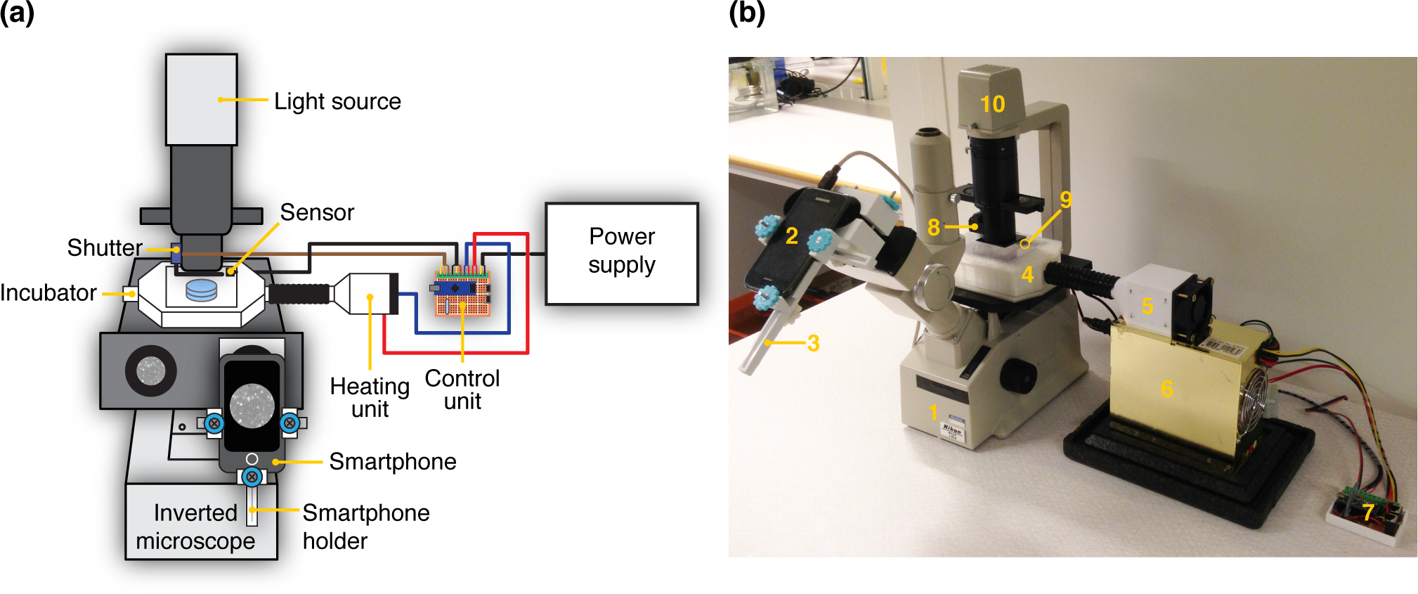
However, in the design of what they call ATLIS (Affordable Time-Lapse Imaging and Incubation System), the researchers managed to create the necessary conditions for a minimal cost. They designed a modular system composed of four main parts: an imaging module, a heating unit, an onstage incubator, and a control unit.
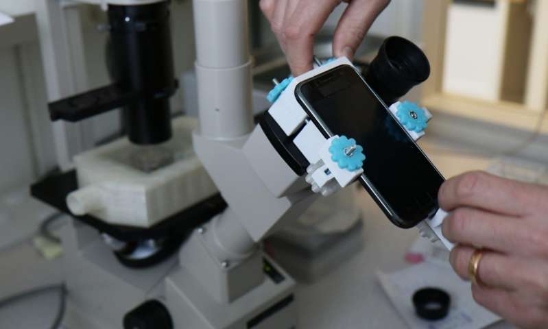
The imaging module was designed to capture high-quality images at a fixed interval using a smartphone camera, while minimizing the amount of light the cells were exposed to. A 3D printed smartphone holder, which was based off of a design found on Thingiverse, was used to attach the phone to one of the microscope’s oculars, as well as to adjust and fix its position in order to capture quality images throughout the duration of the experiment. A shutter was created using a 3D printed connector, a servo motor, and a shutter disc.
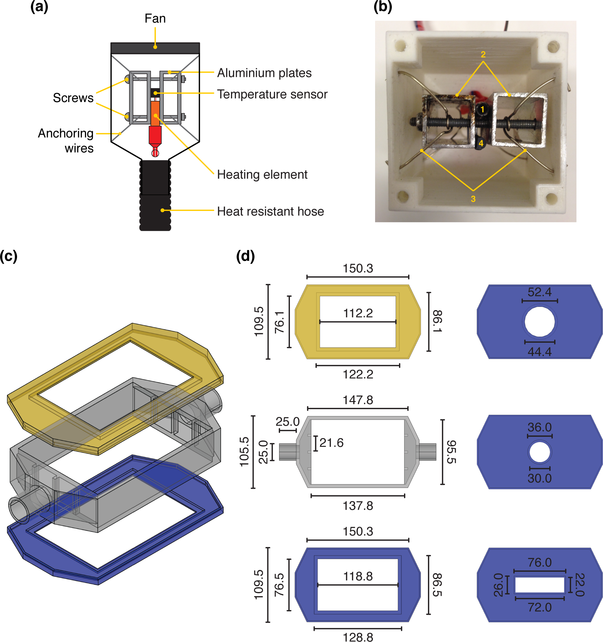 The heating unit was designed to warm the air flowing into the incubator so that the cells stay at an optimal temperature. The core of the heating unit was created by placing a heating element between two commercial-grade square aluminum profiles and held together with two 50mm M4 screws. The core was then placed inside a 3D printed case and suspended with anchoring wires, while a 60mm fan was attached to the back of the case and an air outlet on the opposite end was connected to the inlet of the incubation chamber using a heat-resistant hose. A temperature sensor was placed close to the heating element so that temperature could carefully be monitored.
The onstage incubator was designed as a three-piece system with a lid, a body and a bottom plate, all 3D printed, with a plexiglas-covered opening so that the plate could be exposed to light and imaged. An inlet allowed for warm air to come in from the heating unit, and an outlet allowed air to escape. Four interchangeable bottom plates allowed for different types of cell cultures to be placed inside to be grown, maintained and incubated while imaging took place.
Finally, a control unit was built using an Arduino 3.0 Nano AT328 microcontroller, a Bluetooth communication module, a power transistor and a voltage regulator. The unit was attached to a standard computer supply unit and used to power the other components, while the shutter’s servomotor and the temperature inside the incubator were controlled by Bluetooth.
So how did the ATLIS compare to more high-end equipment? You can read the full study here, but in short, the hacked-together imaging system performed brilliantly. A 12-hour-long imaging session of human embryonic kidney cells showed no evidence of cell death, proving that the environmental conditions created by the ATLIS were fully adequate – plus, the five-megapixel smartphone camera produced high-resolution images comparable to those taken by the specially designed microscope cameras found in expensive commercial live cell imaging equipment.
[Image: Linda Koffmar]
“What we have done in this project isn’t rocket science, but it shows you how 3D-printing will transform the way scientists work around the world. 3D-printing has the potential to give researchers with limited funding access to research methods that were previously too expensive,” said Johan Kreuger, senior lecturer at the Department of Medical Cell Biology at Uppsala University. “The technology presented here can readily be adapted and modified according to the specific need of researchers, at a low cost. Indeed, in the future, it will be much more common that scientists create and modify their own research equipment, and this should greatly propel technology development.”
Other than the smartphone and the microscope, the total cost of the components used to build the ATLIS was $277. All of the 3D printed parts were printed in PLA on a Prusa 3D printer, and the STL files are available upon request. Additional authors of the study, entitled “A Modular and Affordable Time-Lapse Imaging and Incubation System Based on 3D Printed Parts, a Smartphone, and Off-The-Shelf Electronics,” include Rodrigo Hernández Vera, Emil Schwan and Nikos Fatsis-Kavalopoulos. Discuss in the Microscope forum at 3DPB.com.
[Source: Phys.org]
Subscribe to Our Email Newsletter
Stay up-to-date on all the latest news from the 3D printing industry and receive information and offers from third party vendors.
Print Services
Upload your 3D Models and get them printed quickly and efficiently.


You May Also Like
The State of the Desktop Filament Market 2026, Part 1: Facehugging AI
As many as ten million desktop 3D printers may be sold this year. This makes the desktop filament market potentially a significant business. I’ve interviewed key participants in this industry,...
Ugee Releases AI-Powered Funbox 3D Printer for Kids
ugee has released the Funbox, a 3D printer for kids. The company is a part of Hanvon Ugee Technology Group, a manufacturer and distributor of drawing tablets under the ugee,...
Kentstrapper Releases the 1 Cubic Meter Material Extrusion Mille System for €40,000
Kentstrapper makes Material Extrusion systems in Italy. Now the firm has released the 1,000 × 1,000 × 1,000 mm build volume Mille system. The one-cubic-meter build volume is actively heated by...
Pogačar & Fairlight Cycles Show Us Low Cost 3D Printed Components for Bikes
There has been a lot going on in 3D printing for bicycles over the years. The most successful implementation so far is in bicycle seats. Carbon 3D printed seats are...