Researchers Inspired by Pastry Chefs: 3D Printed Constructs Lead to More Successful Tissue Engineering

![]() Looking at the latest project from research at Rice University and the University of Pennsylvania rather broadly, we’d have to say that it’s pretty amazing what you can do with hardware such as a 3D printer and some simple ingredients like sugar and silicone. Just these few resources have been the catalyst for answering a very complex question: How can you deliver oxygen and nutrients to all cells in an artificial organ or tissue implant that takes days or weeks to grow in the lab prior to surgery?
Looking at the latest project from research at Rice University and the University of Pennsylvania rather broadly, we’d have to say that it’s pretty amazing what you can do with hardware such as a 3D printer and some simple ingredients like sugar and silicone. Just these few resources have been the catalyst for answering a very complex question: How can you deliver oxygen and nutrients to all cells in an artificial organ or tissue implant that takes days or weeks to grow in the lab prior to surgery?
It just takes a team of bioengineers and surgeons to put their heads together and figure this stuff out–with that ‘stuff’ being made of cellular material that can be actually be used to grow structures for replacement tissues and organs for transplantation.
 With a John S. Dunn Collaborative Research Award supporting the research, led by Jordan Miller, assistant professor of bioengineering at Rice, and Pavan Atluri, assistant professor of surgery at University of Pennsylvania, this team has so far been able to 3D print an implant with an intricate network of blood vessels.
The researchers have recently released their findings in a study called ‘In vivo anastomosis and perfusion of a 3D printed construct containing microchannel networks,’ published in the journal Tissue Engineering Part C: Methods, and authored by Dr. Renganaden Sooppan, Miss Samantha J. Paulsen, Mr. Jason Han, Mr. Anderson H. Ta, Mr. Patrick V Dinh, Dr. Ann C. Gaffey, Miss Chantel Venkataraman, Mr. Alen Trubelja, Mr. George Hung, Dr. Jordan Miller, and Dr. Pavan Atluri.
Here, they outline how tissue engineering has advanced to such a point, as well as what they now bring to the table in terms of being able to ‘mimic’ vascularized tissue and actually make direct connections between ‘engineered microvascular networks and ‘host vasculature.’
“We have previously demonstrated that the rapid casting of 3D printed sacrificial carbohydrate glass is an expeditious and reliable method of creating scaffolds with 3D microvessel networks,” states the team. “Here, we describe a new surgical technique to directly connect host femoral arteries to patterned microvessel networks.”
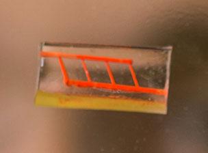
Red dye shows the tiny vessels in the silicone construct created in the Miller lab using a 3D printer. [Photo: Jeff Fitlow]
Previously, those involved in the engineering of tissue would have implanted engineered tissue scaffolds inside the body and waited for them to come alive thanks to adjacent, healthy blood vessels. That process though involves waiting, waiting, and more waiting….and then often, after weeks, the cells die from lack of oxygen anyway.
“We had a theory that maybe we shouldn’t be waiting,” Miller said. “We wondered if there were a way to implant a 3D printed construct where we could connect host arteries directly to the construct and get perfusion immediately. In this study, we are taking the first step toward applying an analogy from transplant surgery to 3-D printed constructs we make in the lab.”
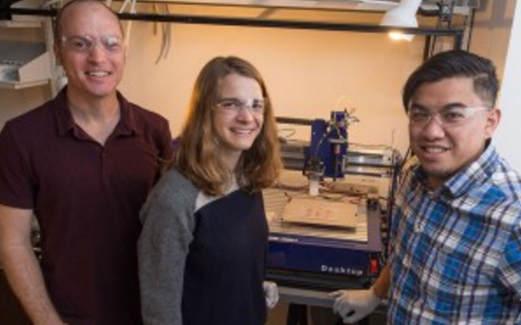
From left, Jordan Miller, Samantha Paulsen and Anderson Ta stand before the 3D printer used to create the silicone constructs. [Photo: Jeff Fitlow]
With this idea, they used the 3D printer to begin fabricating layers of sugar glass, one after the other. This began the lattice network for blood vessels, with the hardened sugar being molded and poured into silicon gel which would then begin to cure. Afterward, when the sugar was dissolved, it left behind a network of small channels in the silicone.
“They don’t yet look like the blood vessels found in organs, but they have some of the key features relevant for a transplant surgeon,” Miller said. “We created a construct that has one inlet and one outlet, which are about one millimeter in diameter, and these main vessels branch into multiple smaller vessels, which are about 600 to 800 microns.”
The hope is that these new research techniques will help overcome the previous challenges met when creating or implanting engineering tissue and then failing to keep it viable, or alive. According to Miller, this study breaks new ground in that they do believe they are much closer to developing a way for surgeons to connect arteries to the tissue that they construct in the lab, ultimately lending success to transplantation. Let us know your thoughts on this research in the 3D Tissue Engineering forum thread on 3DPB.com.
[Source: Rice University News & Media]Subscribe to Our Email Newsletter
Stay up-to-date on all the latest news from the 3D printing industry and receive information and offers from third party vendors.
You May Also Like
3D Printing Unpeeled: New Arkema Material for HP, Saddle and Macro MEMS
A new Arkema material for MJF is said to reduce costs per part by up to 25% and have an 85% reusability ratio. HP 3D HR PA 12 S has been...
3D Printing News Briefs, January 20, 2024: FDM, LPBF, Underwater 3D Printer, Racing, & More
We’re starting off with a process certification in today’s 3D Printing News Briefs, and then moving on to research about solute trapping, laser powder bed fusion, and then moving on...
3D Printing Webinar and Event Roundup: December 3, 2023
We’ve got plenty of events and webinars coming up for you this week! Quickparts is having a Manufacturing Roadshow, America Makes is holding a Member Town Hall, Stratafest makes two...
Formnext 2023 Day Three: Slam Dunk
I’m high—high on trade show. I’ve met numerous new faces and reconnected with old friends, creating an absolutely wonderful atmosphere. The excitement is palpable over several emerging developments. The high...