Now, researchers at Columbia University Medical Center in New York City have developed a new method for treating damaged menisci: 3D-printed implants. Research team leader, Jeremy Mao, DDS, PhD the Edwin S. Robinson Professor of Dentistry (in Orthopedic Surgery) explained why current treatments for meniscal injuries were less than satisfactory. “At present,” explained Dr. Mao, “there’s little that orthopedists can do to regenerate a torn knee meniscus.” The new treatment, however, would allow surgeons to replace the damaged tissue completely.
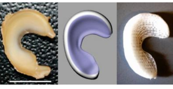
In a paper published on Dec. 9, in Science Translational Medicine, the team at Columbia explained the process, which has been successfully tested in sheep. The procedure begins with MRI 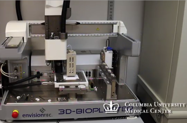
That’s only part of the process, however. In order to ensure that the new meniscus will form from the scaffolding once it is implanted into the patient, the scaffold is infused with connective growth factor (CTGF) and transforming growth factor β3 (TGFβ3), both recombinant human proteins. In a feat of precise tissue engineering, the two proteins are released in very specific order in particular regions of the scaffold. This system of sequential delivery, the Columbia team found, attracts stem cells that already exist in the patient’s body and encourages them to create meniscal tissue.
The scaffold is inserted via a simple surgical procedure into the knee and it takes between four to six weeks for the 3D printed meniscus to regenerate. The scaffold eventually dissolves and the body eliminates it. Typically, with tissue engineering procedures, stem cells are taken from the body and the critical work happens in a lab. In this new approach, the cellular interaction takes place in the patient’s body.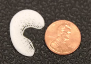
Scott Rodeo, MD, a sports medicine orthopedic surgeon and researcher at the Hospital for Special Surgery in New York City is the co-author of the 3D-printed menisci study. Dr. Rodeo believes that this procedure “would potentially be applicable to the many patients who undergo meniscus removal each year.” So far, sheep have been the test patients because their knees closely resemble those of humans. At this stage in the study, the research team is looking at the endurance of the regenerated tissue. However, the team is optimistic and believes that once the procedure has been refined and approved for use in human patients, the process of getting your new, personalized 3D printed scaffold, once your MRI has been completed, could take a matter of days. The researchers are awaiting funding before they can move on to clinical trials.
Let us know your thoughts on yet another amazing medical use for 3D printing. Discuss in the 3D Printed Meniscus forum thread on 3DPB.com. Check out the video below showing the printing process in action: