
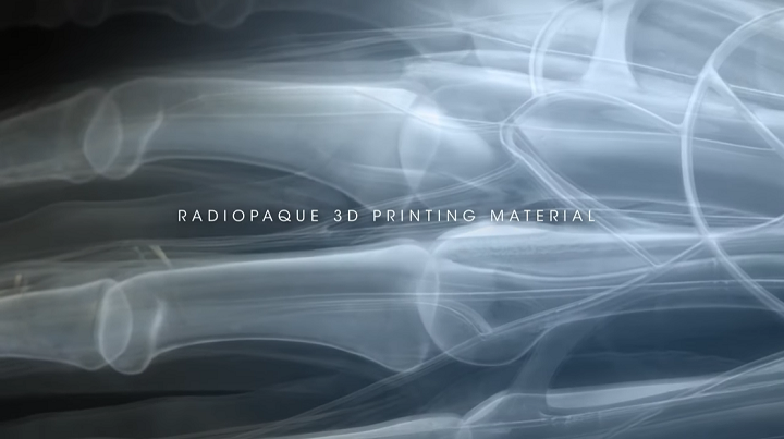
Polymer 3D printing leader Stratasys Ltd. (NASDAQ: SSYS), fresh off reporting what it calls its strongest quarter in six years, has announced the latest 3D printing material for its Digital Anatomy 3D printers, and it’s a doozy. The 3D printing stalwart calls its new Stratasys RadioMatrix material the first radiopaque option for its Digital Anatomy systems, which can be used to print anatomical, radio-realistic models that show up as white or nearly white on X-rays, and are visible under X-ray images or CT scans.
Technically, radiopaque refers to a tissue or material with a bone or near-bone density that blocks the passage of X-rays. According to Stratasys, its new RadioMatrix offers “unprecedented visibility” of 3D printed implants, medical devices, and anatomical structures under medical imaging, which will be a major improvement for surgical training, testing, and navigation. The company says the PolyJet material offers controllable values, repeatable results, and freedom of geometric design.

“This new material is allowing our customers to print radio-realistic models that exhibit defined and predictable radiopacity properties under medical imaging such as CT or X-ray. This material development was driven by customer requests to create radiopaque anatomic models, ones that can mimic human anatomy visualization under X-ray. We continue to push the boundaries of medical modeling with multi-material printing,” Ben Klein, Director of Product Management – Healthcare Solutions for Stratasys, stated in a press release.
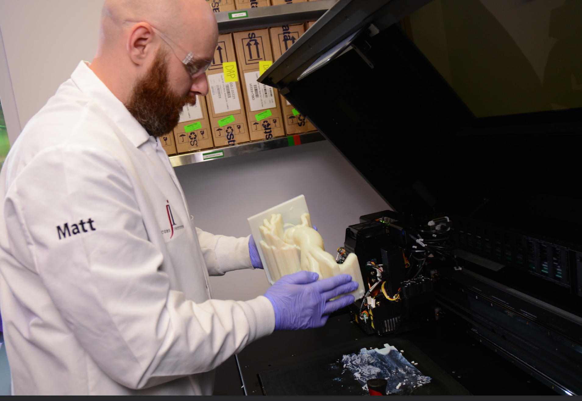
Jacobs Institute expert removing a vascular model from Stratasys J750 Digital Anatomy printer. Image courtesy of Jacobs Institute/Stratasys.
Stratasys first released its Digital Anatomy solution in 2019, which combined custom software and three materials with its multimaterial J750 system to print realistic 3D printed anatomical models for surgical planning and training purposes. The solution was upgraded in 2020 to allow the PolyJet printers to create better bone models, and late last year Stratasys introduced its Digital Anatomy Creator software for easier creation of patient-specific anatomical models.
Using GelMatrix, BoneMatrix, TissueMatrix, and now RadioMatrix PolyJet materials, the Digital Anatomy 3D printers are able to fabricate full-color, functional models that are “biomechanically realistic” and can be used for the aforementioned planning and training applications, in addition to medical device development. Radio-realistic anatomic models printed out of the new RadioMatrix material, in addition to being viewable under CT and X-ray, have a range of radiopacity values from -30 to 1,000 Hounsfield Units (HU).

So far, Stratasys is receiving good reviews for the latest Digital Anatomy material.
“The ability to 3D print models with controlled radiodensity is expected to ultimately improve visibility and traceability of medical devices, improve our production of bespoke and patient-derived phantoms for training and educational purposes, and enable new methodologies for improving CT image quality,” said Justin Ryan, PhD, Director of the Helen and Will Webster Foundation 3D Innovations Lab at Rady Children’s Hospital in San Diego, California. “This novel material opens the door to new applications and research opportunities, ultimately improving patient care delivery.”
The new RadioMatrix material for 3D printed radiopaque anatomical models is already in many regions around the world, and will be coming to the United States sometime in Q3 of this year.
