Chinese researchers are working to improve drug delivery systems for bone repair, detailing their findings in the recently released ‘A novel vehicle-like drug delivery 3D printing scaffold and its applications for a rat femoral bone repairing in vitro and in vivo.’
Bone regeneration continues to be an area of challenge for researchers and doctors as they attempt to treat patients and improve their quality of life overall, performing a wide range of studies regarding materials, properties, and methods. Biomaterials are in great demand, and the authors point out that this demand increases as issues like traffic injuries rise, along with natural disasters and other catastrophes delivering patients in need to hospitals.
With the development of synthetic scaffolds being critical to further progress in bone regeneration, the researchers realize the following properties must play a role:
- Pore architecture
- Mechanical strength
- Bioactivity
- Degradation
- Controllable drug-delivery ability
Porous 3D printed scaffolds are of great interest to researchers, with the authors noting that other studies have centered around bioactive ceramics and bioactive glass of numerous kinds, offering the potential to bond with bone tissue via bioactive materials like hydroxylapatite (HA) or beta-tricalcium phosphate (β-TCP).
Bioglass in particular offers better bioactivity and also releases silicon and calcium ions for better bone regeneration. And while it may have potential for bone tissue grafting, it still lacks the necessary versatility required for drug delivery systems:
“Herein, applying a mesoporous bioactive glass [MBG] coating onto the surface is a viable option for the drug delivering capacity of bioactive glass scaffolds,” stated the researchers. “With mesopores on the surface, DNA, cytokines and some drugs can be loaded onto the scaffold, meeting the requirements of disease treatment or further enhanced bone regeneration.”
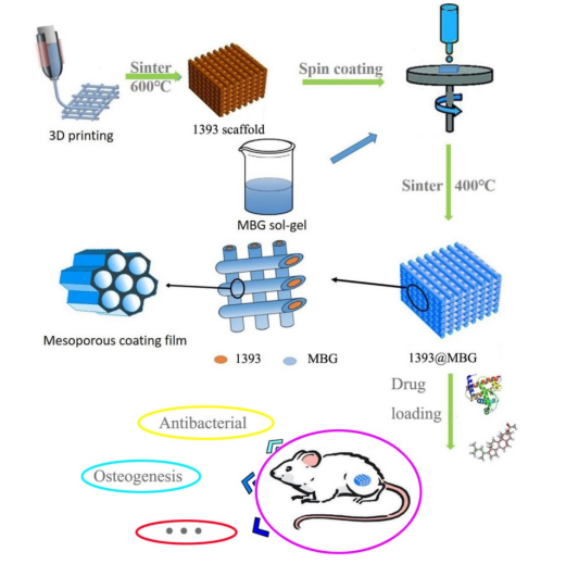
The flow chart of fabricating a novel drug delivery 1393@MBG scaffold built by 3D printing technology for bone repairing.
This study centered around integrating MBG with bioactive glass scaffolds, adding dexamethasone (DEX) and bone morphogenetic protein 2 (BMP-2), imbuing the structures with antibacterial and osteogenic properties. The scaffolds were evaluated for suitable drug delivery, cultured with hBMSCs in vitro and a rat femoral defect model in vivo. After immersion in simulated body fluids (SBF) and staining with ninhydrin, drug release was assessed, testing values at 241 nm for DEX, 260 nm for DNA and 567 nm for BMP-2.
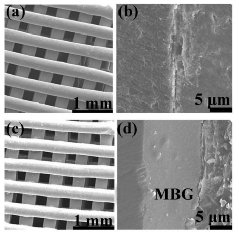
FESEM images of (a, c) as fabricated 1393 and 1393@MBG scaffold; (b, d)
the cross section of as fabricated 1393 and 1393@MBG scaffold.

(a, b) FESEM images and the surface profile of as fabricated 1393 scaffold; (c, d) after immersed scaffold; (e, f) FESEM image and the surface profile of as fabricated 1393@MBG scaffold; (g, h) after immersed scaffold; (i) The Ra of 1393 and 1393@MBG scaffold surface when immersed from 0 to 90 days; (j) The compressive strength of the 1393 and 1393@MBG scaffold on day 0, 30 and 90. mean ± SD, n = 5. *Significant difference when compared to 1393 (p < 0.05).
The study reflected positive results, with MBG proving the capacity to both load and control drug release. BMP-2 exhibited the most rapid rate of release, followed by DNA, and then DEX.
“The main group of MBG was silicon oxide group, thus when immersed in a water solution the surface of MBG will exhibit a positive charge which will more easily absorb negative materials such as –OH groups, DEX and DNA,” stated the authors. “So, this may explain the phenomenon that BMP-2 release was better than that of DNA and DEX, in the order, BMP-2 > DNA > DEX.”
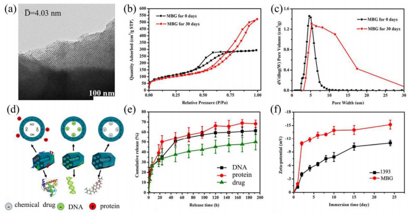
(a) TEM image of the as made MBG powder; (b) N2 adsorption–desorption isotherms and (c) the corresponding pore size distributions of MBG powders; (d) illustrator of the mechanism of MBG loading protein, DNA and chemical drug; (e) chemical drug, DNA and protein release profiles from the MBG powders in SBF at 37 ℃; (f) Zeta-potential of 1393 and MBG glass powder immersed in SBF at 37 °C as a function of immersion time; mean ± SD, n = 5.
“We have investigated the response to the functional scaffolds of hBMSCs in vitro and the osteogenic capacity in rat femoral defects in vivo,” concluded the research team. “Results showed that the coating of MBG on 1393 scaffolds were a viable way to enhance the proliferation and ALP activity of hBMSCs and upregulate osteogenesis-related genes expression. The prepared BMP-2 loaded 1393@MBG scaffolds significantly improved bone regeneration in the osseous defects at 12 weeks post-implantation. These results suggested that the novel drug delivery 1393@MBG scaffolds could be a promising candidate for the use in bone tissue repair and relative disease treatment.”
What do you think of this news? Let us know your thoughts! Join the discussion of this and other 3D printing topics at 3DPrintBoard.com.

The attachment of hBMSCs on the BMP-2 loaded 1393 scaffolds (a) and 1393@MBG scaffolds (b) after culturing for 2 days; The live (green)/dead (red) staining for the 1393 (c) and 1393@MBG scaffold (d) immersion solution.