
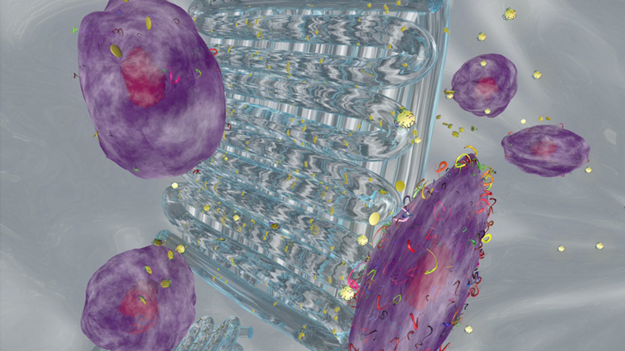
In the paper Printing Therapeutic Proteins in 3D using Nanoengineered Bioink to Control and Direct Cell Migration published by the team last May, they propose using the high surface area and charged characteristics of 2D nanosilicates (or nanoclays, nanoparticles of layered mineral silicates) to sequester and deliver biologically active therapeutics. Therapeutic proteins such as vascular endothelial growth factor (VEGF) and fibroblast growth factor (FGF) were incorporated into the bioink for in vitro testing, which was then used
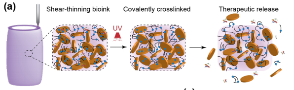
The nanoengineered bioink loaded with therapeutic proteins is based on a nanoclay platform pioneered by Gaharwar that can be used for precise deposition of protein therapeutics, which Gaharwar developed as a new way to deliver treatment for cartilage regeneration and to eventually impact the treatment of osteoarthritis. This bioink formulation has unique shear-thinning properties with high printability and structural fidelity, that allow the material to be injected, quickly stop flowing and then cure to stay in place.
Charles Peak, senior author of the study and Assistant Instructional Professor at the Department of Biomedical Engineering at Texas A&M, said that “the prolonged delivery of the bioactive therapeutic could improve cell migration within 3D printed scaffolds and can help in rapid vascularization of scaffolds.”
Synthesis and characterization of PEGDTT/nanosilicate bioink loaded with protein therapeutics
While team leader, Gaharwar, said that the prolonged delivery of the therapeutic could also reduce overall costs by decreasing the therapeutic concentration as well as minimizing the negative side effects associated with supraphysiological doses (amounts greater than normally found in the body).
“Overall, this study provides proof of principle to print protein therapeutics in 3D that can be used to control and direct cell functions,” he suggested.

PEGDTT bioink by itself remained fluid, while PEG-DTT/nanosilicate can be printed in 3D constructs with high mechanical stability
According to the published results, the shear-thinning bioink with the ability to sequester and release therapeutic proteins within 3D printed scaffolds is developed from a hydrolytically degradable polymer and 2D synthetic nanoparticles, with strong mechanical properties, swelling kinetics, and a degradation rate that can be modulated via the amount of PEG-DA and nanosilicates. The activity of released protein therapeutics from the 3D structure was verified via the migration of cells.
Supported by the National Institute of Biomedical Imaging and Bioengineering of the National Institutes of Health and the National Science Foundation, the research is in the midst of emerging bioprinting technologies for regenerative medicine and methods for rapidly fabricating
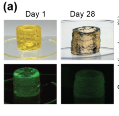
Bioink loaded with protein therapeutics: the release of fluorescently labeled protein from 3D printed structure was monitored over 28 days
cell-containing constructs for designing new, healthy, functional tissues. According to Texas A&M, one of the major challenges in 3D bioprinting is the lack of control over cellular functions. Growth factors, which are a special class of proteins, can direct cellular fate and functions. However, these growth factors cannot be easily incorporated within a 3D-printed structure for a prolonged duration, which is why these developments at their Department of Biomedical Engineering lab are so relevant. And with so many papers on this subject, we will probably hear more about the team’s work with bioinks.
[Images: Texas A&M, Wiley Online Library]