Researchers are using 3D printing for nerve reconstruction, learning more about how nerves work both functionally and internally in a peripheral capacity. They outlined their methods and results in the recently published ‘An enhanced staining method K-B-2R staining for three-dimensional nerve reconstruction.’
In using the 2D staining method with Karnovsky–Roots toluidine blue ponceau 2R (and then toluidine blue counterstain and ponceau 2R counterstain), the researchers were able to ‘significantly improve the ability to display nerve fascicles, motor, and sensory nerve fiber textures.’ These nerves don’t fit the general definition of ‘peripheral,’ as they offer critical functions in receiving impulses and sending out instructions. With 3D technology, researchers can see incredible intricacies as nerves branch out and connect. They can also learn more about disease and trauma.
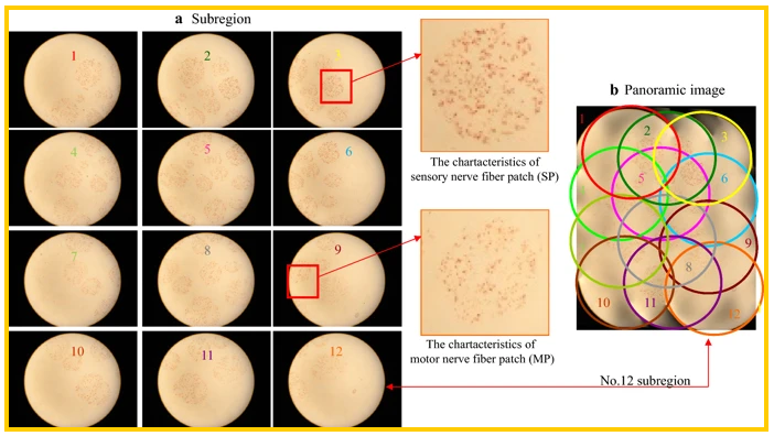
The 12 sub-region images of a median nerve section (No. 20) following Karnovsky–Roots staining and their mergence to create the panoramic image, a sub-region and b panoramic image
Current nerve reconstruction with 3D technology is based on the acetylcholinesterase histochemical method, preparing:
- Specimens
- Acetylcholinesterase staining
- 2D image acquisition
- 2D image processing
- 3D reconstruction
Current procedures with Karnovsky–Roots stained images can be arduous, and accuracy is unpredictable; in fact, in many cases, the nerve fascicle regions and sensory nerve fiber textures may not be visible. This makes it difficult to reconstruct the nerves or create a suitable model. Here, the team worked to refine traditional methods, creating a way for better visibility of the nerves and fibers.
“First, Karnovsky–Roots staining was conducted, and subsequently toluidine blue counterstaining was performed, followed by ponceau 2R counterstaining,” stated the researchers. “This K-B-2R staining procedure was found to be able to better display the microstructure of myelin sheath, enhance the textural property of ROI, elevate the degree of recognition of section images and facilitate image partition and 3D nerve reconstruction and 3D printing.”
The nerves were designed with Mimics, and then 3D printed with the Raise3D N2 Plus 3D printer, with PLA as the chosen material. Better recognition of the nerve allows for better recognition overall, image partition, and accomplishment.
“In addition, the 3D printing technology was applied to create the 3D digital model of nerve fascicle. Thus, this new staining technique can facilitate 3D reconstruction and creation of the 3D digital model, which suggests that this new technique can facilitate to rebuild and repair the nerve fascicles when it is used in conjunction with the 3D reconstruction and 3D printing technologies,” stated the researchers.
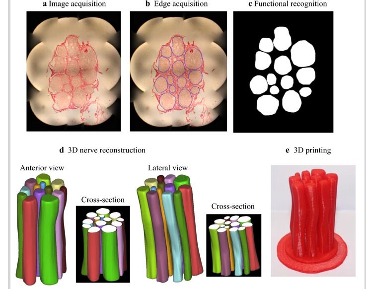
Staining, functional recognition, 3D reconstruction and 3D printing results of another segment of the median nerve (No. 40) after K-B-2R staining. a Image acquisition; b edge acquisition; c functional recognition; d 3D nerve reconstruction (front view and lateral view); and e 3D printing
The researchers found that with this method, it took much less time for processing and adjustment, and partition results were almost just like true nerve fascicles—making the technique better over alternative approaches.
“This 2D K-B-2R staining method has significantly shortened the cycle and evidently enhanced the precision of 3D peripheral nerve reconstruction. This technique has thus appropriately resolved the technical challenge faced by nerve injury repair,” concluded the researchers. “Furthermore, the 3D reconstruction and 3D printing technology can provide an ideal solution for the nerve injury repair in the field of nerve tissue engineering if the appropriate printing material is applied.”
3D printing is useful in many different types of reconstruction and regeneration for the human anatomy, making enormous impacts on the medical realm—and ultimately, on patients’ lives, whether due to scaffolding created for bone regeneration, mandibular reconstruction, methods for fabricating ears, or more. Find out more about nerve reconstruction here. What do you think of this news? Let us know your thoughts! Join the discussion of this and other 3D printing topics at 3DPrintBoard.com.
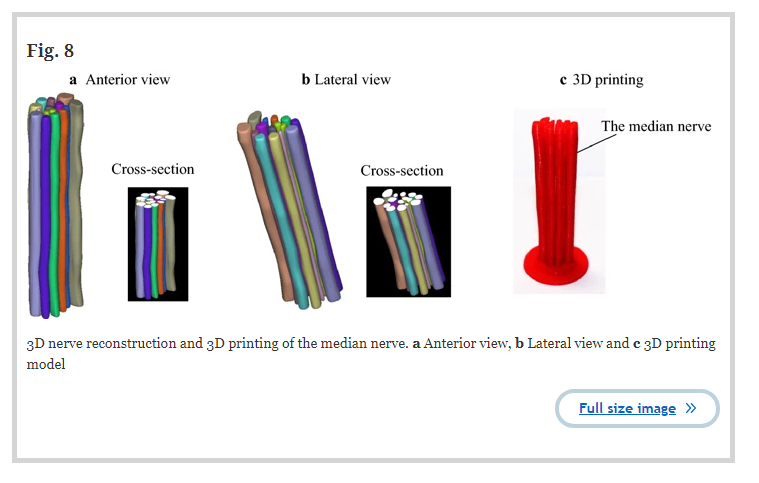
3D nerve reconstruction and 3D printing of the median nerve. a Anterior view, b Lateral view and c 3D printing model