This resource is the first of its kind, catering to the patient-specific 3D modeling requirements for the Mount Sinai clinicians. The simulation, prototyping, and 3D printing resources developed at Mount Sinai are pretty rare for a medical institution. The 3D printed models will be used in the planning stages for minimally invasive approaches, and can also be used in a surgery trial run. They will also be invaluable during the patient consultation process. Surgery can be a pretty scary thing, and patients who can see and touch these models will surely feel more at ease before their procedures take place.
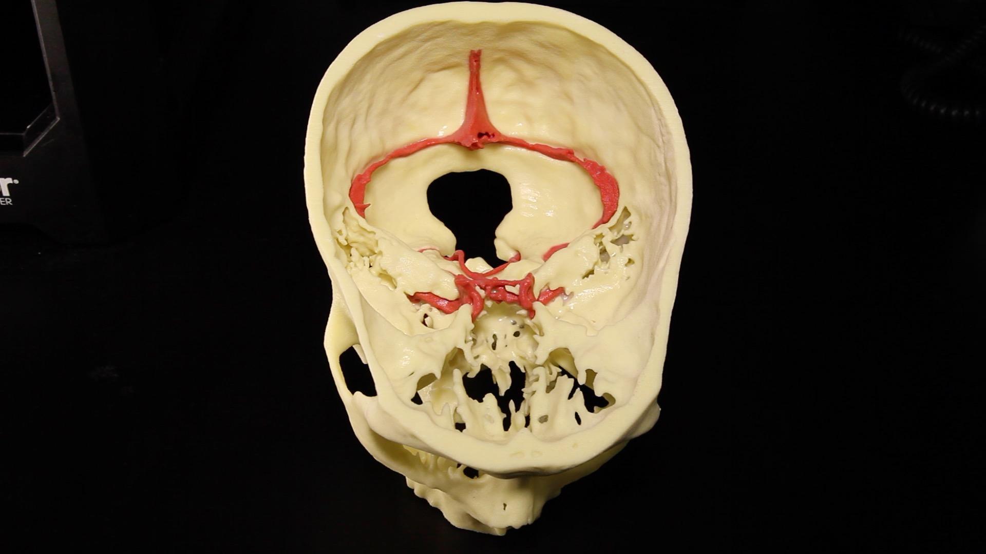
Multi-structure 3D print of patient-specific pathology including epidermoid cyst (green), arterial vasculature (red), fifth cranial nerve (orange), seventh and eighth cranial nerves (yellow), and skull base (white). Each component of the print was individually segmented from registered CT and MR data to produce the final result. Multicolor printing was achieved using a 3D Systems ProJet 660 3D Printer. [Image/Caption: ISMMS Neurosurgery]
Anthony Costa, PhD, Assistant Professor for the Department of Neurosurgery and Scientific Director of the Neurosurgery Simulation Core at the Icahn School of Medicine at Mount Sinai (ISMMS), is leading the 3D printing team. He has developed segmentation tools and computer code that will accelerate the process of turning radiological data into 3D printable models. Some of the recent 3D prints completed, according to Mount Sinai, “include skull-base tumors with surrounding vasculature and cranial nerves, spine modeling for the correction of severe scoliosis, and pelvic models for the planning of arthroplasty” (the surgical repair of a joint using tissue). Several interdisciplinary collaborations have already been established between the new Medical Modeling Core and Mount Sinai clinical departments, including cardiology, surgery, orthopedics, and otolaryngology (the branch of medicine that deals with the ear, nose, and throat).
“We’re unique because we can leverage our technological tools with the expertise of radiology and the printing lab to complete projects on a rapid time scale,” said Dr. Costa. “We’re talking about days as opposed to weeks. Mount Sinai is a large institution with a high volume of cases and our patients will benefit from 3D modeling.”
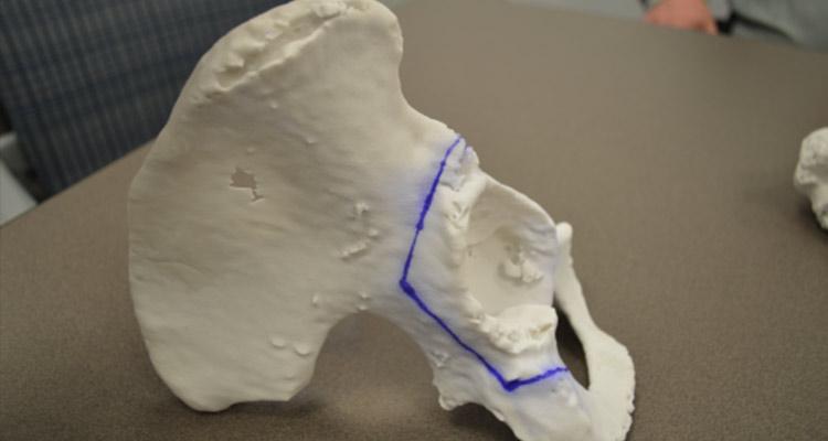
3D print of pelvis component for surgical planning, with blue outline highlighting targets for surgical approach. [Image: ISMMS Neurosurgery]
As an example of how these 3D services work, the neurosurgery department collaborates with the Department of Radiology at the ISMMS, to ensure that the best quality imaging is done for the 3D simulation. During the generation of 3D data, structures in the head that are important for a specific patient’s pathology, are identified through the radiological imaging and ‘segmented’ from their surroundings. For example, with a brain tumor, the brain’s vascular network and skull are modeled, along with the cranial nerves. This segmentation results in a set of models that accurately depict the relationship between structures of the brain that are critical for planning the operation. After this, the Surgical Theater’s Surgical Navigation Advanced Platform (SNAP) is typically used to show both the patient and the surgeon this pre-op data.
[Image: Mount Sinai]
The design and production of the 3D models is done in-house, which is also a significant cost-saving measure for the physicians at Mount Sinai. A $500 3D printed model created at the hospital could represent a ten-fold savings over having outsourced the model. The Rapid Prototyping Center utilizes four 3D printers, and a laser cutter, to produce high resolution, patient-specific neuroanatomy for pre-operative planning. Among available printing materials are polyamide (nylon), epoxy resin, wax, and polycarbonate.
Mount Sinai Health System includes approximately 7,100 primary and specialty care physicians, 31 affiliated community health centers, and is ranked No. 15 nationally in the 2016-2017 “Best Hospitals” issue of US News & World Report. The ISMMS is ranked among the highest in the nation in National Institutes of Health funding per investigator.
To learn more about the new Medical Modeling Core, watch this video: