It can be due to abnormal development of the bones of the forehead and the base of the skull, can occur due to premature fusion of the bony plates of the skull, or can be caused by a variety of syndromes, such as Edwards, duplication, basal cell nevus, DiGeorge, and Loeys-Dietz syndromes, among other potential causes.

Dr. Satish Vasishta
Enter Osteo3D, a Bangalore, India-based company backed by the df3d design factory, which specializes in 3D printing solutions for the healthcare field.
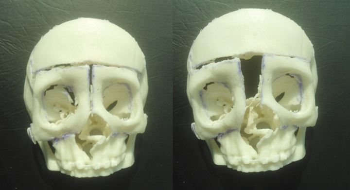
Dr. Sathish Vasishta, a craniomaxillofacial surgeon, and Dr. Derick Mendonca, a consulting plastic surgeon, used a 3D printed model to successfully plan and perform a surgical procedure at Sakra Hospital in Bangalore, which used a pair of techniques called ‘Box Osteotomy’ and ‘Facial Bi-Partition’ to take on the problem.
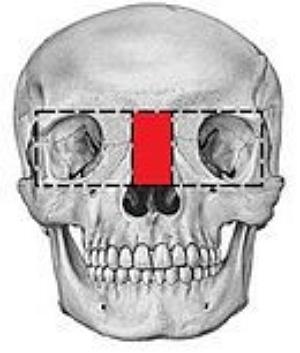
The doctors say the patient, Esther, was suffering from excess bone which had grown between the orbits. To correct the condition, it was necessary to remove bone on either side of the midline of the skull. And to complicate matters further, the patient also showed signs of what’s known as Orbital Dystopia, a condition where one bony orbit is not symmetrical with the other.
According to the surgical team, using the model also helped them prevent mistakes during the operative procedure and allowed them to correctly design the “osteotomies” which would correct the Orbital Hypertelorism.
Before they entered the operating theater, the surgical team rehearsed the work by actually practicing the procedure on the 3D printed model. They say the pre-operative session allowed the doctors to accurately estimate what it would take to remove some 7.5 mm of bone on either side of skull’s midline in the horizontal plane and an additional 1.5mm of bone in vertical plane of the skull surrounding the right bony orbit.
By using the techniques they developed in the rehearsal session, and moving the bony compartment of the orbit to the midline on the model, the doctors achieved what’s called Orbital Symmetry for the patient on both the horizontal and vertical planes of the skull.
Do you have any other stories we should know about where 3D printing technology has been used to help patients and doctors? Let us know in the Hypertelorism With 3D Printed Modeling forum thread on 3DPB.com.