While 3D printing offers many users the ability to create and innovate completely new designs and products, such technology can also be put to very good use for filling in the blanks. This can be true in terms of industrial components that may have become obsolete and need to be recreated for vehicles, assault rifles, or vessels, parts of the human body that need to be rebuilt and fitted with implants after severe trauma, or as in this latest research shows—anthropological finds and skeletal remains that need to be reconstructed, whether in law enforcement or scientific settings.
In “Effective approaches to three-dimensional digital reconstruction of fragmented human skeletal remains using laser surface scanning,” authors from India and the UK explore 3D printing for remodeling skeletal parts that may be missing. For this study, they focused on remodeling part of a human cranium and reconstructing a mandible. Historically, forensic anthropologists have used superimposition with photography for identifying missing people—but have experienced challenges in rebuilding due to areas where the bone is lost, and parts of the cranium, for example, may be compromised.
Today, analysis of human remains relies on methods like:
- 3D imaging
- X-rays
- Micro CTs
- Laser scanning
- Structured-light scanning
- 3D photogrammetry
3D digitization has offered clear advantages in many projects over the years, commonly in museum or research settings, allowing for affordable replicas that can be used in scientific exchange, criminal forensics, teaching, museum displays, and more. The benefits to forensic departments continue worldwide, as well as contributing to work performed by other professionals.
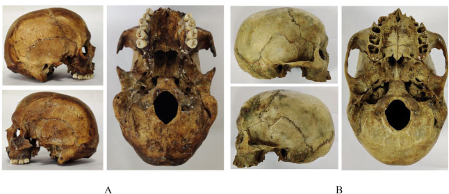
A: Lateral and basal view of cranium with bilaterally missing zygomatic process; B: Lateral and basal view of cranium with intact zygomatic process.
The researchers were able to obtain two human craniums from the Gujarat Forensic Sciences University skeletal archives for 3D scanning using a NextEngine 3D Laser Scanner, Geomagic Studio 13 for surface reproduction, and a Flashforge Guider 2 3D printer.
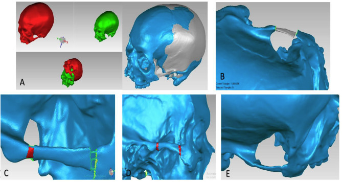
Steps for reconstruction of missing zygomatic process (A) Manual Alignment and Global Registration (B) Replacing the missing element in the test data after extracting from reference data (C) & (D) Bridging the gaps using various methods. (E) Smoothening.
A cranium model was 3D printed with PLA, measuring 280 × 250 × 300 mm.
“A direct articulation was established between zygomatic process of maxilla and temporal bone after reconstruction of zygomatic arch,” stated the researchers. “The results were confluent with that mentioned in literature.”
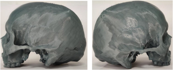
3D printed reconstructed model.
A human mandible was also obtained via Gujarat Forensic Sciences University. The replica was made with vulcanized silicone and then filled with dental stone. The researchers broke the replica into nine pieces.
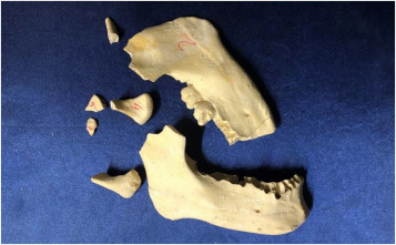
Fragmented replicated mandible.
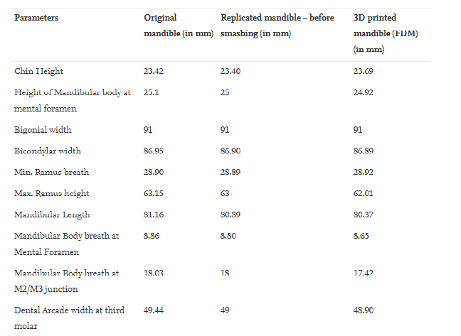
The resulting measurements verified that the 3D printed model could be used for “various morphometric analysis.”
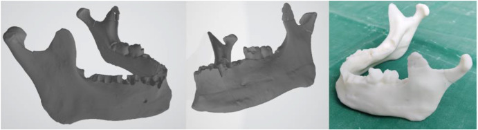
Conversion in ‘.stl’ file after aligning all the parts (left) and printing using FDM technology (right).
Many different projects involving rebuilding of bones rely on scanning of the good side or an intact area and then mirroring the incomplete data. In this study, the incomplete zygomatic arch was created using data from a similar cranium. The researchers concluded that such a model would be suitable for use in court, as well as in forensic anthropology and medicine.
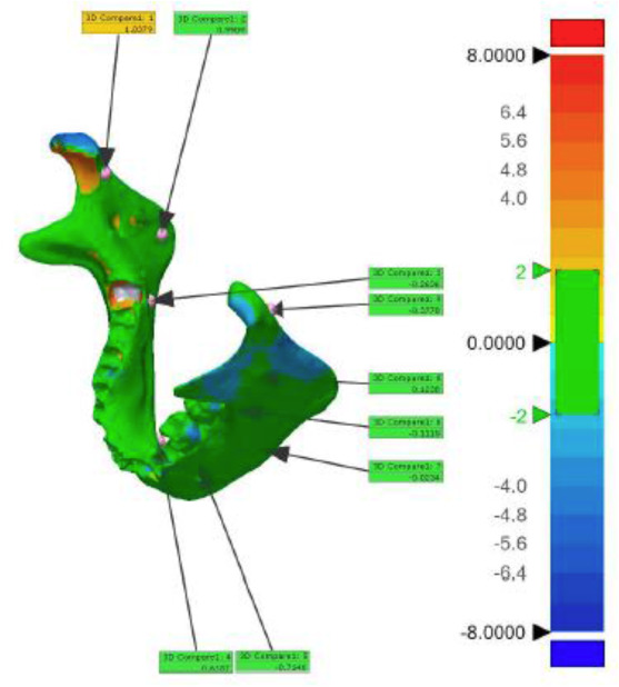
Qualitative congruency analysis performed on scanned data of a reference mandible and a reconstructed mandible.