Chinese researchers are exploring ways to treat patients with osteoporosis, releasing the findings of their recent study in ‘Enhanced osseointegration of three-dimensional supramolecular bioactive interface through osteoporotic microenvironment regulation.’
As researchers continue to investigate a variety of ways to use scaffolding in tissue engineering, from new methods to new materials offering regrowth and regeneration, for this study the scientists centered around designing a prosthetic surface interface for solving issues with complications due to osteoporosis.
Such a prosthesis has the potential to solve some of the more severe issues faced after joint replacement surgery, to include prosthesis interfacial displacement, loosening, and fracture. Titanium alloy is commonly used for orthopedic implants due to resistance to corrosion and overall strength; however, the authors note that obstacles are presented in its use due to high stiffness, often resulting in osteolysis (pathological resorption of bone matrix).
While 3D printed porous titanium alloy (pTi) scaffolds offer a host of benefits, there is the potential for failure due to inferior integration into the bone. To overcome such issues, the research team experimented with injecting supramolecular hydrogel, with BMSCs and BMP-2 dual-loading, into the inner pores of 3D printing porous metal scaffolds.
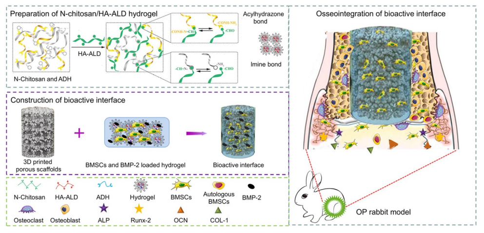
Ti filled with BMSCs and BMP-2 dual-loaded supramolecular hydrogels as bioactive composite scaffolds for enhancing osteoporotic bone defect osseointegration. BMP-2 can promote osteogenic differentiation of exogenous BMSCs and endogenous BMSCs. With the degradation of hydrogel, bone tissue grows into the pores of the pTi scaffold, thus achieving good osseointegration.
Spherical pre-alloyed medical-grade Ti6Al4V powder was used to make the scaffolds via EBM 3D printing:
“Disk-shaped scaffolds (Ø10 mm × L3 mm) were used for microstructural and cellular biocompatibility and osteogenic assays in vitro (titanium plates without porosity were printed for the control group in vitro cell experiments), and the columnar-shaped scaffolds (Ø6 mm × L10 mm) were used for mechanical testing and in vivo osseointegration investigations,” the researchers wrote.
“Concisely, hydrogel was prepared by in situ crosslinking of N-chitosan and ADH with HA-ALD; 7.5% N-chitosan (w/v) and 7.5% ADH (w/v) were dissolved in deionized water. Next, solutions of 5% HA-ALD (w/v) were added to the above mixture. For hydrogel formation by imine and acylhydrazone bonds, the solution was stirred using Lab Dancer to obtain a homogeneous hydrogel.”
The researchers then performed a topography characterization of the bioactive interface, mechanical characterization of the bioactive interface, in vivo degradation and biocompatibility analysis, and other morphology and examination.

(A) Bioactive interface constructed by BMSCs and BMP-2 loaded hydrogel filled the pTi in a cell culture plate; (B) Representative optical image and 3D micro-CT image of pTi; (C) Mixed hydrogel and gelation; SEM images of (D) pTi, (E) supramolecular hydrogel, and (F) pTi filled with hydrogel.
Porosity was in order with the initial design, at 70.5±0.9%, with the sol-to-gel transformation and mechanism ‘attributed to acylhydrazone and imine bonds.’ SEM images were captured, along with the Image J analysis, showing pTi scaffold pore sizes at 793.4±16.9 μm.
“Therefore, the porosity and pore size of pTi scaffold in this study were suitable for bone tissue engineering. Increasing the surface area and porosity of the scaffolds could improve the initial implant stability, bone ingrowth capacity, and the friction coefficient between the bone and scaffolds, thus reducing micromotion and promoting osseointegration after implanting in vivo,” stated the researchers.
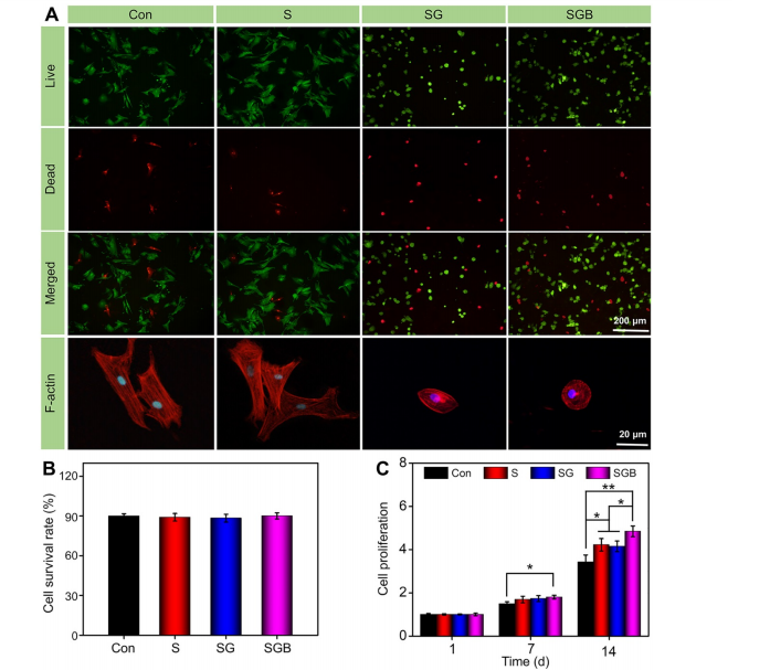
(A) Calcein AM/PI staining of live cells (green) and dead cells (red), and fluorescent imaging with rhodamine-DAPI staining of Con, S, SG, and SGB groups; (B) Quantitative analysis of cell survival rate by Calcein AM/PI staining; (C) Cell proliferation within the different scaffolds at 1, 7, and 14 d (*p < 0.05, **p < 0.01).
The hydrogel was degraded within one month, leaving the research team to note that there was no significant inflammation, and space was available for bone growth within the pTi.
“This composite system was demonstrated possessing good biocompatibility, ensured the sustained release of bioactive BMP-2, and was beneficial for osteogenic differentiation of BMSCs. As a synergic therapy, BMSCs and BMP-2 dual-loaded hydrogel could induce bone ingrowth and promote osseointegration of microporous titanium in osteoporotic bone defects,” concluded the researchers.
“These findings suggested that this bioactive interface was a potentially promising candidate for the development of the artificial prosthesis interface for various osteogenesis-deficient patients, such as osteoporosis and rheumatoid arthritis.”
What do you think of this news? Let us know your thoughts; join the discussion of this and other 3D printing topics at 3DPrintBoard.com.
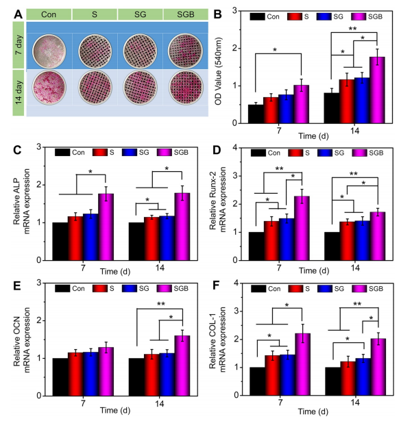
Evaluation of the osteogenic differentiation of BMSCs seeding in the Con, S, SG, and SGB groups. (A) Images of different samples after staining with alizarin red S; (B) Statistical analysis of semi-quantitative analysis of alizarin red staining; (C–F) The expressions of osteogenic-related genes, such as ALP, RUNX-2, OCN, and COL-1 (*p < 0.05, **p < 0.01).