Chinese researchers investigate the benefits of using biodegradable polymers for scaffolds, outlined in ‘Fabrication and characterization of porous polycaprolactone scaffold via extrusion-based cryogenic 3D printing for tissue engineering.’ Explaining that there have historically been limitations due to issues like affordability, lack of efficiency in fabrication, and inferior process control, the authors of the study endeavor to improve on previous attempts to use 3D porous PCL scaffolds through combining extrusion-based cryogenic 3D printing with freeze-drying approaches.
Tissue engineering (TE) is a broad field today and one that is expansive with research and many different goals—most of which end in the ultimate discovery of a way to create sustainable bioprinted organs for transplantation. In creating or regenerating tissue, scientists usually work with scaffolds, living cells, and other ‘bioactive factors.’ Structures like scaffolds must be biocompatible, and obviously non-toxic too if they are being implanted into a human patient. Polycaprolactone (PCL) is a commonly used polymer in creating scaffolds, suitable due to features like:
- Biodegradability
- Biocompatibility
- Low melting point
- Good strength
- Good solubility
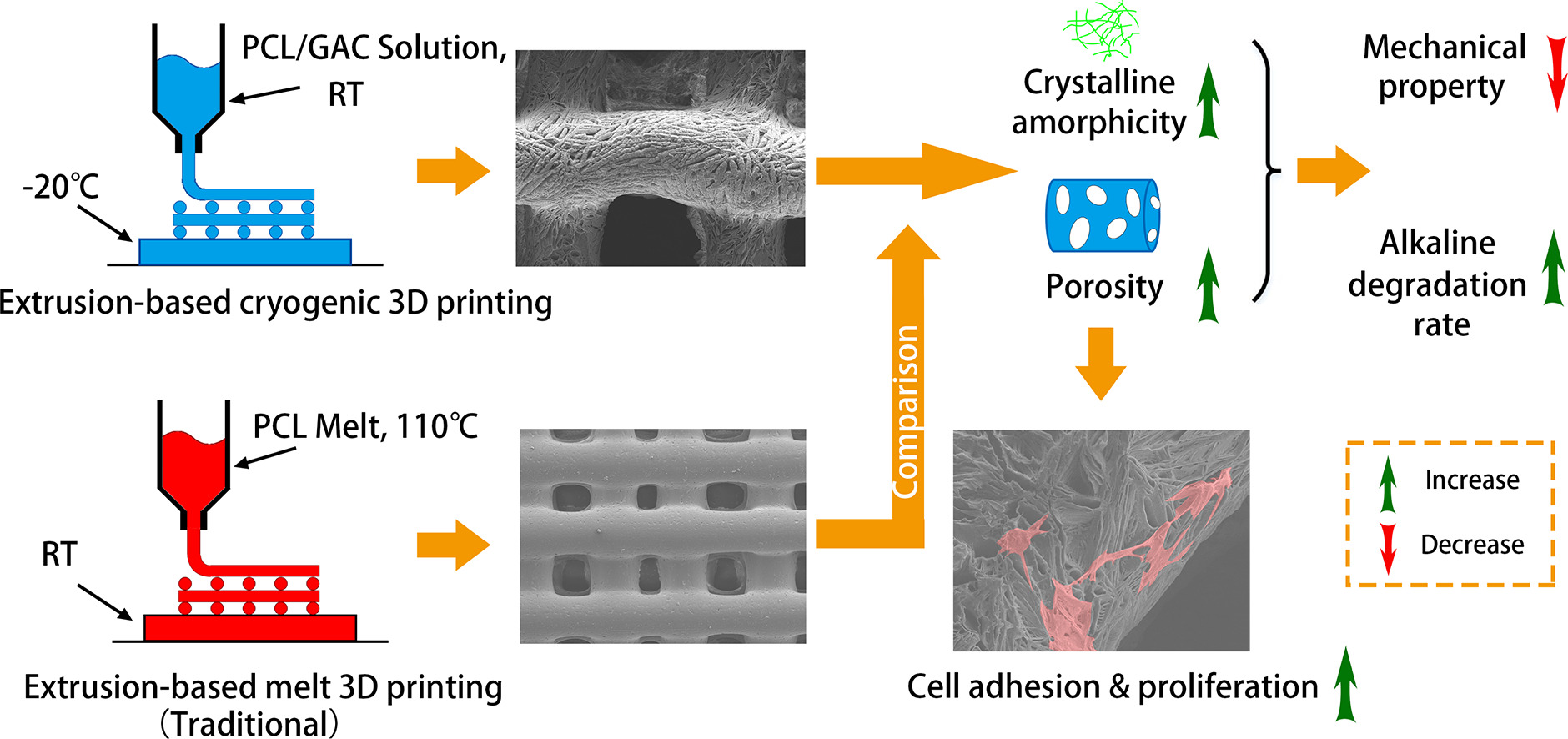
The researchers explain that extrusion-based cryogenic 3D printing (ECP) is gaining more popularity as a choice for bioprinting because it allows for greater strength in scaffolds, whether they are made of collagen, chitosan, PLGA, or other materials. In this study, the authors used ECP to fabricate PCL scaffolds and then study the results.
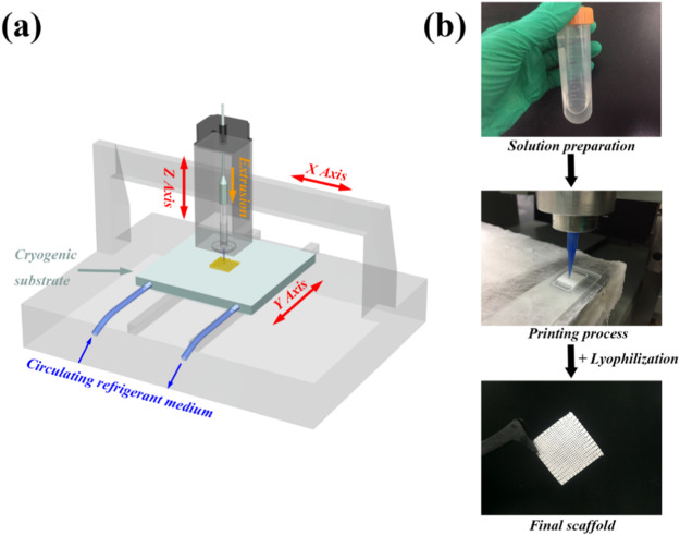
(a) Schematic illustration of a cryogenic 3D printing platform. (b) Pictorial representation of extrusion-based cryogenic 3D printing and lyophilization of PCL scaffold.
To ensure success in printing, the researchers relied on several different treatments, to include using a rough-surfaced glass slide as a collector, adding a transitional path at the corners of the adhesive area, and scrubbing slides with ethanol, to begin with. Porosity was measured, with results showing an increase due to ‘widening of filament offset.’
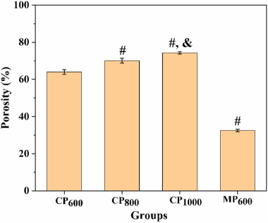
Porosity analysis of printed scaffolds. (# denotes groups comparing with CP600 at P < 0.005; & means a comparison between CP800 and CP1000 at P < 0.05).
In terms of measuring biocompatibility, the researchers found that while cell attachment was ‘not well promoted’ at first, cell proliferation was ‘effectively facilitated’ because of the rough surface and porosity of scaffolds.
“Although more stretched cells were found on the surface of EMP group after 7 days, the number of cells on ECP scaffolds were much higher and their morphologies become more stretched as compared to the ones at day 3,” concluded the researchers. “Thus, it can be concluded that PCL scaffolds fabricated via ECP are highly biocompatible and better support cell adhesion and proliferation as compared to EMP scaffolds.
“Overall, the fabricated PCL scaffold, with such improved structural, physico-chemical, and biological features, can be a promising candidate for tissue engineering applications.”
Tissue engineering takes many forms today, from heart tissue engineering to bone tissue engineering to tailored skin grafts. What do you think of this news? Let us know your thoughts! Join the discussion of this and other 3D printing topics at 3DPrintBoard.com.
[Source / Images: ‘Fabrication and characterization of porous polycaprolactone scaffold via extrusion-based cryogenic 3D printing for tissue engineering’]