![]()
Just as traditional 3D printing is performed with filaments—most often made from a variety of different colorful polymers—bioprinting is performed through different types of cell-laden bioinks. Different types of hardware may be preferred too, to include:
- Inkjet bioprinting – an affordable method often created through droplet generation featuring piezo-electric voltage.
- Stereolithography – also known as SLA, this method employs laser technology and curing that relies on polymer solutions (filled in a reservoir) for materials.
- Laser-induced forward transfer (LIFT) – this system is comprised of a laser, focusing apparatus, and ribbon responding to light, with the bioink layer usually maintained under the donor layer.
- Extrusion – these types of systems are most common for bioprinting, with bioink loaded into cartridges and then extruded through nozzles.
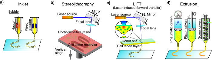
Schematic graph of the most commonly employed bioprinting techniques. (a) Inkjet printers use a heat or piezo actuator to generate small drops, which are collected at an underneath platform. (b) Stereolithography (SLA) printers use the movements of a laser beam to crosslink a photosensitive polymer solution into a planar pattern, and a 3D structure is formed by vertical accumulation of the patterns at each layer. (c) Laser-induced forward transfer (LIFT) printers use an energy absorbing material stimulated by a light source to generate drops of the donor material, and the drops are collected and constructed into 3D structures at the underneath platform. (d) Extrusion-based printers extrude soft materials out of nozzles by pneumatic, piston, or screw driven actuation and form 3D structures by the vertical accumulation of each layer.
The researchers go into much detail regarding extrusion-based bioprinting and its advantages such as the ability to create materials dense with cells, along with allowing the creation of heterogeneous models—and as the authors point out, this is particularly vital to tissue engineering and the future engineering of human organs. Extrusion bioprinting has also become more affordable over time and is extremely customizable—a facet that is usually enticing to experienced users, and especially in the science lab. As for disadvantages, sustainability of cells can be a major issue—and both resolution and speed may still be somewhat inferior in many systems too.
“Current efforts have been focused towards the design and optimization of bioinks and implementation of better extrusion mechanisms, nozzle diameters, and control systems, to increase printing resolution and achieve better deposition times without compromising cell viability and model fidelity,” state the researchers.
Three different categories of bioprinting have been achieved so far, to include bioprinting of biomimetic matrix structures optimized for subsequent cell-seeding, bioinks that support the direct deposition of embedded living biological matter, and engineering bioactivity and biofunctionality into bioink formulations. Recent popularity in bioprinting has also sparked off commercialization of the technique and accompanying systems. The authors also point out numerous ‘practical considerations’ regarding bioprinting:
- Bioink can be challenging to maintain and is prone to dehydration.
- Surface tension may minimize surface energies of bioink, affecting the entire system—and structures being printed.
- Thermal diffusion may be uneven within the bioink cartridge, damaging sensitive materials.
- Cell viability is a constant point of concern as it can be exceedingly difficult to keep cells alive for successful bioprinting.
Soft matter bioinks are discussed also, with hydrogels leading the way as the main topic. This is an extremely popular vehicle today for bioprinted structures and can retain large amounts of water within their networks. The researchers point out that there are many advantages to using hydrogels, but they also may lack the proper anchoring required for cells to adhere and then migrate.
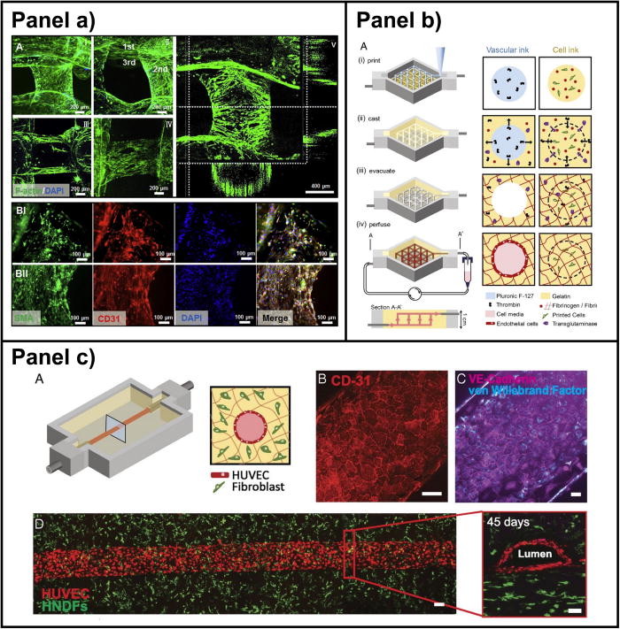
Vascularized 3D bioprinted models. Panel (a) is printed tubes composed of GelMA/alginate/PEGTA hydrogel containing endothelial and stem cells using a coaxial nozzle device.97 Reproduced with permission from Jia et al., Biomaterials 106, 58–68 (2016). Copyright 2016 Elsevier Ltd. Panel (b) is a schematic illustration of thick vascularized tissue within 3D perfusion silicon chips using gelatin and fibrinogen as bioinks. Reproduced with permission from Kolesky et al., Proc. Natl. Acad. Sci. U. S. A. 113(12), 3179–3184 (2016). Copyright 2016 National Academy of Sciences. Panel (c) shows a vascularized 3D printed tissue comprised of HUVEC-lined vascular channel surrounded by fibroblast-laden matrix and casted within a 3D perfusion chip.19 Reproduced with permission from Kolesky et al., Proc. Natl. Acad. Sci. U. S. A. 113(12), 3179–3184 (2016). Copyright 2016 National Academy of Sciences.
These types of bioinks have led to the creation of many complex models today meant to promote tissue regeneration—from vascular models to those containing brain tissue. Challenges persist though—and as is usually true with any new technological development at first, a lack of standards; in this case, standards would be relative to printability. The number of cells in bio-ink is another challenge. A bioink may be cell-laden, but in comparison to what? And what types of nozzle and settings will cause the least amount of stress on cells? The last consideration the researchers ponder is whether bioprinting really possesses the ability to imitate human tissue as required; however scientists are making rapid strides in eliminating obstacles to bioprinting as the stakes for changing the face of medicine as we know it are so very high.
“As stated, the technique is rapidly developing, and we firmly believe that the currently existing challenges can be addressed in the future,” conclude the researchers.
It is hard to describe one element of 3D printing that is ‘taking the world by storm’ these days as there are so many innovations continually being presented to the world—meant for a variety of powerful applications. Bioprinting is significant, however, as researchers are not only engineering tissue but edging closer and closer to the creation of human organs in the lab. In the meantime, we have followed stories on machine learning and drop-on-demand techniques, creating neural tissue with chitosan-gelatin hydrogels, and even the use of stem cells from Alzheimer’s patients to assist in further research.
What do you think of this news? Let us know your thoughts! Join the discussion of this and other 3D printing topics at 3DPrintBoard.com.
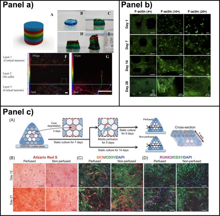
Bioprinted models for tissue regeneration. Panel (a) shows a handheld method of bioprinting used to create 3D brain-like structures composed of primary cortical neurons embedded in a gellan gum.261 Reproduced with permission from Lozano et al., Biomaterials 67, 264–273 (2015). Copyright 2015 Elsevier Ltd. Panel (b) represents a 3D printed model for cartilage regeneration using human nasal chondrocyte (hNC)-laden nanofibrillated cellulose (NFC)/alginate hydrogels.263 Reproduced with permission from Martínez-Ávila et al., Bioprinting 1–2, 22–35 (2016). Copyright 2016 Elsevier Ltd. Panel (c) is a 3D printed bone vascularized tissue construct consisting of degradable GelMA, surrounded by nanoplatelet-loaded GelMA bioink and covered by chemically conjugated vascular endothelial growth factor (VEGF).264 Reproduced with permission from Batzaya et al., Adv. Healthcare Mater. 6(16), 1700015 (2017). Copyright 2017 Wiley-VCH Verlag GmbH & Co. KGaA, Weinheim.