
Pre-operative CT scan depicting the massive tumor in the left pelvis
Chondromyxoid fibroma, or CMF, is a rare, benign bone tumor that’s typically found in the bones of legs, arms, feet, hands, fingers, and toes and occurs most often between the ages of 10 and 30. A few years ago, 18-year-old Noor Fadil was experiencing severe pain in her left hip, which was caused by CMF of the pelvis, and was unable to bear weight on the hip or walk normally, which was obviously causing some issues in her daily life. After one medical facility advised a hindquarter amputation due to the severity of the tumor, she sought a second opinion from the Yellow Ribbon team in Bangalore, India.
“We are a group of orthopaedic oncosurgeons involved in managing Sarcomas,” Dr. Pramod Chinder told 3DPrint.com about the team. “We use 3D Prints regularly Mainly for planning and making guided Jigs.”

Once Fadil’s CMF diagnosis was confirmed, Dr. Chinder, along with fellow Yellow Ribbon team members Dr. Chandramouli, Dr Suraj, and Dr Srinath, collaborated with Bangalore-based Osteo3D and implantcast GmbH in Germany to make a custom 3D printed pelvic implant for Fadil…the first ever in the country, in fact. It’s no easy feat to reconstruct or resect a pelvis, which is why the team turned to 3D printing to get the job done.
“In this era of evolving relationship between technology and medicine, revolutionizing health care; our prior experience with 3D printed models and its application for surgical practice helped us to move a step ahead in utilizing this technology,” the team wrote in about the case.
“Collaboration of medicine and technology is becoming the antidote for issues that formerly caused despair, for both the patient and the surgeon. We feel the need for a generation of physicians and surgeons, who are technologically skilled and adapted toward innovation.”
During a multidisciplinary tumor board meeting, the team planned their approach, starting with using a minimally invasive approach to debulk the lesion in the patient’s pelvis, then using an ultrasonic probe to find “a well-defined cavity.”
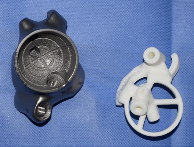
Custom 3D printed hip implant and jig
The surgeons completed removed the tumor, and completed an excision of the head of the femur for good measure, to ensure that no tumor tissue was left. Then Fadil went home and began therapy both to maintain her limb function and to begin forming a strong, healthy bone bed – an essential foundation for a reconstructed hip joint.
Then the waiting began. To make sure she was completely ready for her hip reconstruction, the Yellow Ribbon team followed up with Fadil for two years to make sure that there no signs of the tumor coming back; luckily, there weren’t any. However, the patient’s remaining pelvis was distorted after her surgery, so the team took CT and MRI scans of the area in order to create a realistic digital model for planning purposes and to help design a biocompatible, patient-specific, 3D printed implant.
It was not an easy procedure, because the 3D printed implant needed to be placed at a very specific location, with specific angles, for it to fit properly. The screws to anchor the implant were also well-designed, as they too needed to end up in a certain place since Fadil only had a limited amount of native pelvic bone left. The team designed and used 3D printed plastic guidance jigs to make sure everything ended up where it was supposed to be.
“We operated the child for 8 hrs plus in 3 sittings,” Dr. Chinder told us, noting that Fadil and the team spent a grand total of 24 hours in the operating room. “We are very happy with overall outcome.”
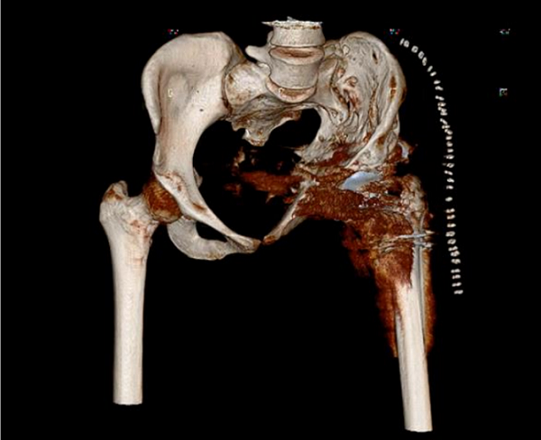
Post-operative CT scan of the pelvis
Post-op, Fadil needed to take things slow, and with plenty of assistance, began non-weight bearing hip range of motion therapy and strengthening exercises during the first week after her surgery. Two months later, she began working on partial weight bearing, and eight months out, she was able to walk normally, bearing full weight, on her left side without relying on any supports like crutches and with no limp.
Just think – if the Yellow Ribbon team hadn’t been there, Fadil may have had to deal with an amputation. This is yet another example of 3D printing being used to change someone’s life for the better.
Discuss this news and other 3D printing topics at 3DPrintBoard.com or share your thoughts in the Facebook comments below.
[Images provided by Dr. Pramod Chinder]