![]()
Nate Yamane was born in June 2015 and had trouble breathing shortly after birth. It turned out that Nate had tetralogy of Fallot (TOF) with pulmonary atresia – the genetic abnormality caused an obstruction to his pulmonary artery, so blood pumped by the heart was prevented from flowing into his lungs. He was taken to CHLA in critical condition. Pulmonary atresia is a more severe form of TOF, and about one in 10,000 children are born this this defect. It occurs when the pulmonary artery does not form properly in utero. To combat this, the body grows “collateral arteries” that will redirect blood flow to the lungs and bypass the obstruction.
“Imagine blood flowing in the artery like cars on the freeway, and it’s blocked,” explained Pediatric Interventional Cardiologist Dr. Frank Ing, Chief of the Division of Cardiology and Co-Director of the Heart Institute at CHLA. “Cars exit and find an alternate route to its destination; blood does the same, and in this case finds its way through collateral vessels to the lungs.”
Nate had a catheterization procedure and two open heart surgeries by the time he was a month old, and that wasn’t even the end of it. Six months into his young life, doctors discovered that Nate’s pulmonary arteries, in the left and right branches, were narrowed. Dr. Ing led a team that was able to open up the right branch using a balloon, but it wasn’t that easy to open up the left. Doctors had to insert a special stent into the narrowed section of Nate’s left pulmonary artery, which at the time was about 15 mm. Stents normally don’t come that small, but Dr. Ing was able to develop a technique to modify it to fit Nate.
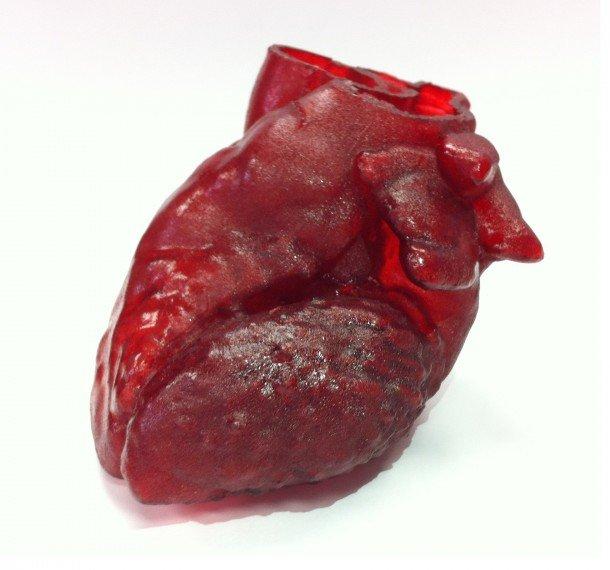
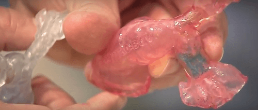
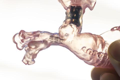
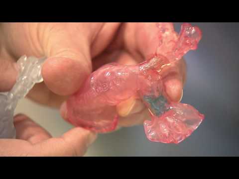
In order to get the measurements exactly right before the procedure, doctors created a 3D printed model of the obstructed region of Nate’s pulmonary artery, using CT scans of his heart, to use as a guide. Dr. Ing was then able to make a custom, fully functional stent by cutting the smallest stent they had and folding it back on itself.
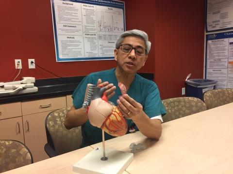
Dr. Frank Ing, Chief of the Division of Cardiology and Co-Director of the Heart Institute at Children’s Hospital Los Angeles [Photo: Children’s Hospital Los Angeles]
Dr. Ing said, “I have to say, the 3-D model was very helpful because it gave me confidence that [the size of the stent] was going to work.”
His fix worked: the stent fit exactly into the narrowed artery in the model, and when they inserted it into Nate’s actual artery, it was perfect, and didn’t jut out into other areas. Dr. Ing saw almost immediate improvement in Nate’s blood flow and a healthy drop in his blood pressure. But later, CHLA cardiologists discovered that a portion of Nate’s right artery was narrowing again, and it was determined that a stent would also be needed to keep that artery open. They ran into the same issue as before: due to the narrowed size (about 9 mm), they needed to create another custom stent to fit into the small opening. But Nate needed to grow a little bigger and stronger first, before the doctors would attempt a second procedure; his mother, Courtney, said they did some “physical therapy and tried to fatten him up” first.
[Photo: Children’s Hospital Los Angeles]
“He’s rolling around with energy and even took his first baby steps. There’s a big difference and a lot of improvement. We’re going in the right direction,” said Courtney.
Take a look at the video of the live feed from Nate’s second stent surgery last month:
Discuss in the 3D Printed Heart Model forum at 3DPB.com.
[Source/Images: Heart Institute at CHLA]