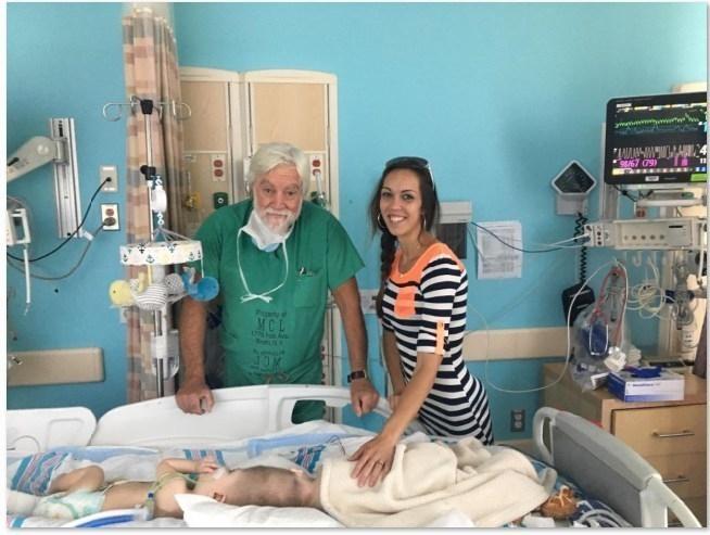
Dr. James Goodrich and Nicole McDonald with the twins prior to separation.
Conjoined twins are extremely rare, and often don’t survive long past birth. The parents of those who do survive are faced with a choice: risk having the children separated in a surgery that could be fatal to one or both, or allow them to remain attached and face serious challenges in functioning throughout life? It’s become safer and easier to separate many types of conjoined twins than it used to be, but in some cases, depending on where the babies’ bodies are fused, it’s still incredibly difficult and dangerous.
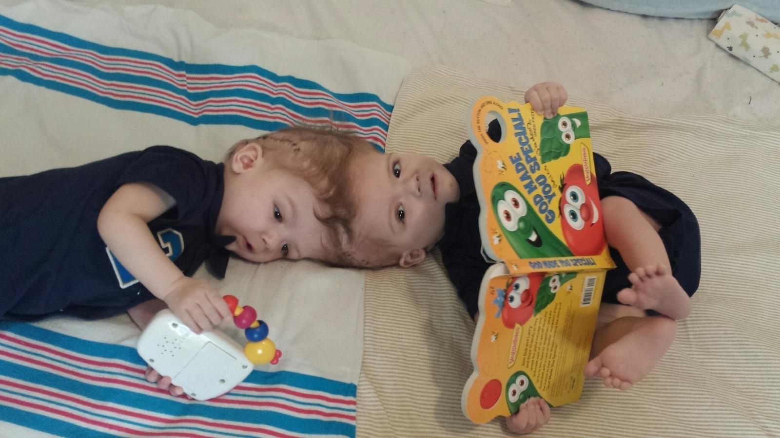
In the case of craniopagus twins, who are joined at the head, the challenges are especially great. Craniopagus twins, if not separated, typically don’t live past their second birthdays, so surgically separating is a necessity – though an extremely dangerous one. Thirteen-month-old craniopagus twins Jadon and Anias McDonald presented a huge challenge to surgeons at the Children’s Hospital of Montefiore (CHAM), as their brains were connected in an extremely complex way.
“This was unexpectedly one of the hardest cases I have ever worked on,” said James T. Goodrich , M.D., Ph.D., D.Sci. (Hon.), director, Pediatric Neurosurgery, CHAM and professor, Clinical Neurological Surgery, Pediatrics and Plastic and Reconstructive Surgery, Albert Einstein College of Medicine. “We knew they shared an area of fused brain, but we did not know how complicated it would be until we looked inside.”
It was thanks to 3D technology that Dr. Goodrich and his team were able to look inside the boys’ brains before operating. Using a combination of 3D printed models and virtual planning software, the surgical team was able to carefully study the connected tissue and plan the course they would take to separate the babies. Even with the preparation, though, the process was far from simple; Jadon and Anias had to undergo four surgeries over the course of seven months before they were finally separated.
In the last decade, the standard procedure for the separation of craniopagus twins has been “staged separation,” or multiple surgeries. Prior to 2004, surgeons would attempt to separate the babies in one long surgery, which was very often fatal to one or both, until it was discovered that doing the procedure in multiple surgeries would allow the children’s veins and vascular systems to recover in the interim.
Dr. Goodrich is one of the leading surgeons in the separation of craniopagus twins; he performed the last such procedure at Montefiore in 2004, separating Carl and Clarence Aguirre, who recently turned 14. He has since consulted on 20 cases of craniopagus twins, and Jadon and Anias are the seventh set that Dr. Goodrich has successfully separated.
“Once again the Montefiore Einstein team is leading the way in pioneering medicine that changes lives,” said Steven M. Safyer M.D., President and CEO, Montefiore. “This intricate procedure was greatly enhanced by cutting edge 3D technology that enabled the surgeons to see inside the boys’ brains. But what really makes this case so special to me is the way our entire community is embracing the McDonald family. This is Montefiore at its best: sophisticated science, medicine and patient care are at the heart and soul of what we do.”
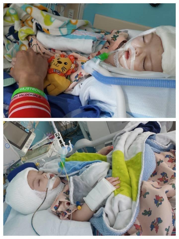
The boys may eventually need additional surgeries, and they have a long way to go towards recovery. They will undergo rehabilitation therapy once they are strong enough, and will eventually go home with their mother Nicole, father Christian, and older brother Aza. The family, originally from Chicago, moved to the Bronx this year for the babies’ surgeries.
![]()