
The ultrasound is a breathtaking experience for parents-to-be, and especially in the first weeks. While you can’t make out much at that point, there is one small but extremely important body part you can both see and hear: the heart. I must say, even today, when my young daughter is cuddled up next to me, settled in and sleepy, I can often feel her heart beating, and I’m taken right back to that moment when the ultrasound technician first located her heart on the screen and showed it to me. It’s the most literal sign of life, and beyond that all that, a parent wants to know that the budding life and heart are also okay—and if not, a thorough explanation is certainly required.

Continued learning lies at this heart of this matter, but for a 3D science, the models need to reflect that in the best way possible, leading to a superior ultrasound reading. And while we’ve followed so many stories regarding 3D printed models helping in complex surgeries such as that of jaw reconstruction and even saving a spleen, this context provides the perfect example of how the models are so valuable just in training, for everyone.

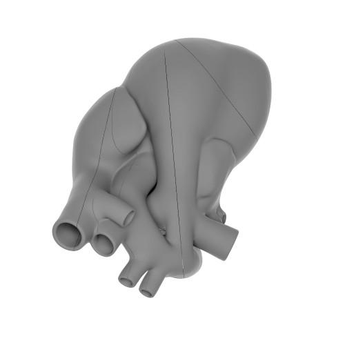
“These models are an important tool in the education of obstetricians-gynecologists and physicians interested in prenatal ultrasonography,” states MWU.
Each model is 10 x 14 x 10 cm, and includes instructions as well as illustrations detailing each layer of the model. There are over 40 different displays to choose from, with some showing how a proper heart should look, along with many examples of different defects emerging at birth.
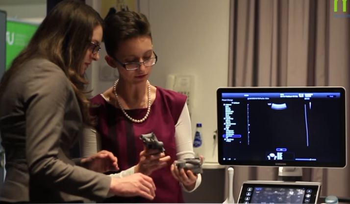
“I was skeptical at first,” said Dr. Agnieszka Nocun, MWU co-founder and lecturer. “But at my visit at GRID when I saw their professional approach and how the model was built from the very basics, A to Z, I agreed with Marcin [Wiechec, co-founder] that it was a really great idea, and our models constitute a great form of educational tool for physicians.”

Exploring the 3D printed fetal hearts at the premiere of the MWU workshop series.
While this is a perfect start for extending education further in the field of ultrasound technology, 3D printed medical models continue to help advance the medical field—with some hospitals and medical facilities even creating entire lending facilities, like the 3D Printed Heart Library at Jump Trading Center in Illinois. All of these models mean so much more than just a piece of plastic representing a human organ. They can be customized to look exactly like a patient’s heart—or brain, or kidney, or liver—and can be shown with a condition or tumor just as it appears in the body, allowing for diagnostics, treatment, and navigation in surgery.
“Our models also had proven themselves during patient consultation,” states the MWU team. “Among others during our workshop in March 2016, we detected a heart defect during practical classes in one of the patients.”
With the 3D printed fetal hearts, everyone involved is able to be better educated, as well as informed on how to move forward when there is a potential issue or defect involved. As the technology continues to evolve, so will the models, in a variety of different intricate designs and materials. If a picture is worth a thousand words, then certainly one of these customized cardiac models is worth hours of training via the chalkboard. Discuss the use of these medical models further over in the 3D Printed Fetal Heart Models forum at 3DPB.com.