Of all the vital areas where 3D printing technology has had an effect, the medical industry has probably shown the most positive and immediate return. Doctors from around the world are increasingly utilizing patient-specific 3D printed models to help with complex surgical preparation, which has led to faster and more effective medical operations. This particularly rings true for what may be considered the human body’s most important organ, the heart. From Toronto to Melbourne, cardiologists and surgeons worldwide have found 3D printed models of hearts and arteries to be the optimal preparation tool. Even at my alma mater, the University of Central Florida, Professor Dr. Dinender Singla has been working valiantly to make these 3D printed heart models more accessible to all.
![]()
Considering the age and condition of their patient, the Queen Elizabeth Hospital team aimed to perform a surgery with minimal invasiveness, and were able to do so thanks to 3D printing technology. The patient-specific model enabled the medical team to optimize their planning and practice, which helped them to complete the procedure in just four hours. The specialists agreed that the best surgical method for Shum would be through her blood vessels, which only requires a small incision on the body for them to access the targeted area.
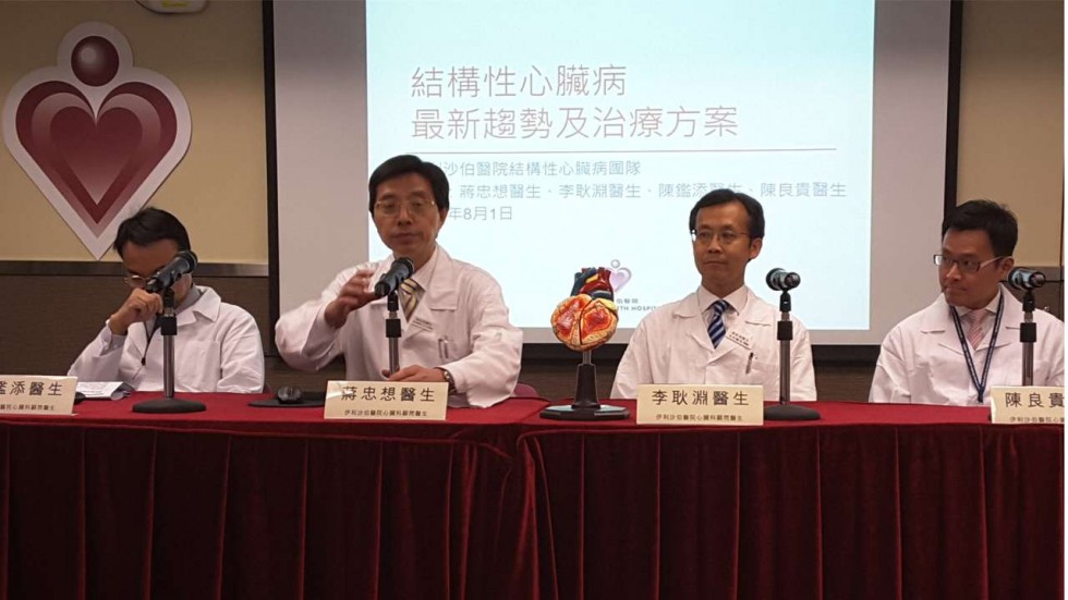
Heart disease specialists from the Queen Elizabeth Hospital [Photo: Shirley Zhao, SCMP]
The patient, who has undergone three open heart surgeries since 1973, had two damaged heart valves, the mitral valve on the left side and the tricuspid valve on the right, leaving Shum at high risk of heart failure. So, the medical specialists decided that another open heart surgery would be precarious, and instead opted to insert a new artificial valve through a minimally invasive opening in a main vein on her upper thigh. The valve was constructed from the heart tissue of a cow placed upon a metal wire, which guided the valve from her thigh all the way up to her heart.
The impressive success of the procedure has helped the Queen Elizabeth Hospital’s structural heart disease division obtain extra funding from the Hospital Authority, which has enabled them to expand their staff. With this, the team hopes to continue enhancing their ability to perform more minimally invasive heart surgeries, which 3D printing technology will almost surely play a major role in. Discuss further in the 3D Printed Heart Model forum over at 3DPB.com.
[Source: South China Morning Post]