Clinical nurse specialist Kerr Clapperton using Artec Spider to scan Ellie’s ear in Artec Studio
Undergoing even a routine medical procedure can be unnerving, no matter your age. But for children, whose understanding of rules and confidence in strangers is just developing, it can be even more terrifying. When a child is born with a condition that requires medical attention, they may become accustomed to the fear, but many medical providers are looking for ways in which that fear itself can be largely eliminated from the interaction.
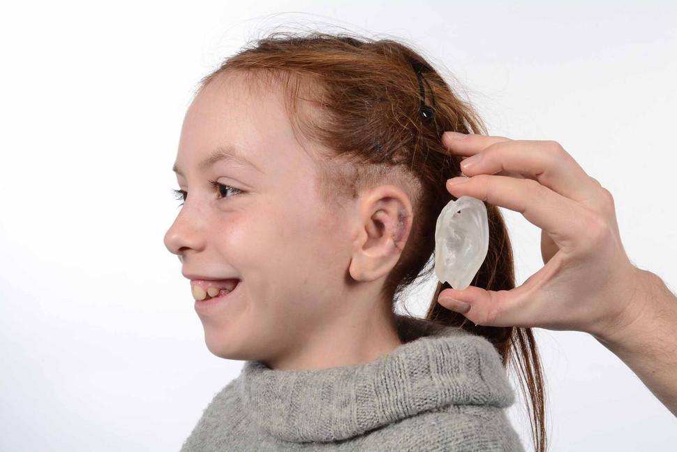
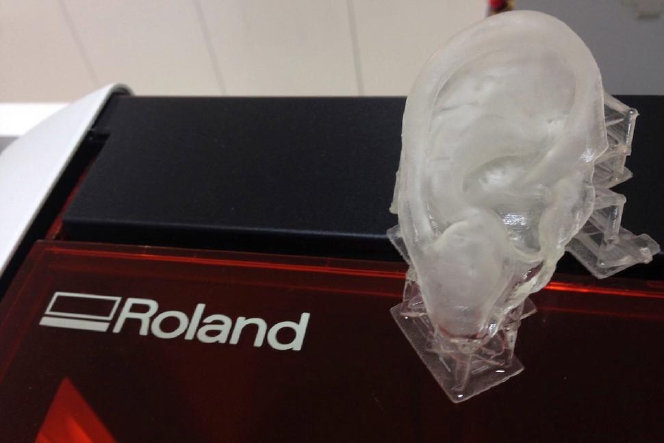
Now, using a handheld Artec Spider 3D scanner, the doctor can record the geometry of the child’s normal ear and use that to create a highly accurate model. No fear, no fuss. This technique was pioneered by Dr. Ken Stewart of the Royal Hospital for Sick Children in Edinburgh, Scotland who recently used it to help a young girl named Ellie, and explained the process in an interview with Digital Trends:
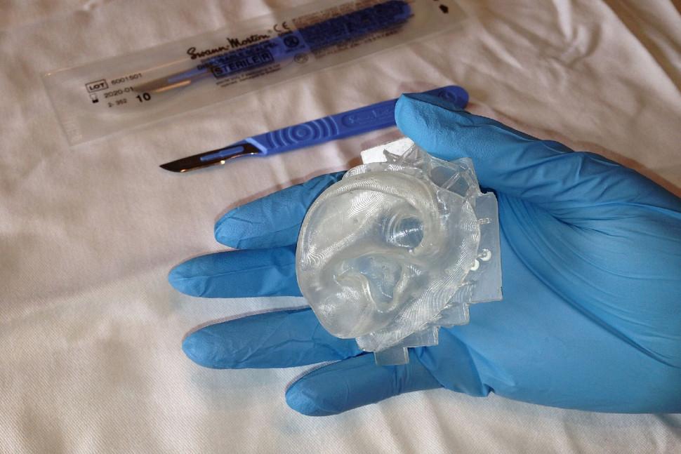
“Most patients are born with one ear missing or a loose part of one ear. Traditionally we would use a clear acetate to take a 2D tracing of the normal ear. This would be used as a template to help carve an appropriately sized and shaped opposite ear. The 3D scanner and mirror image software allows us to produce a more accurate template.”
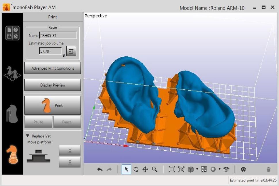
“Professor Bruno Peault and his team have characterized stem cells within human fat which lie next to blood vessels. We can harvest these very easily by liposuction. For a plastic surgeon that is easier than taking blood. With Professor Mark Bradley’s team in the chemistry department we have identified FDA approved polymers to which the stem cells will bind and be driven to produce cartilage. We know we can 3D print in the polymers concerned. So the Artec-derived 3D scans could potentially be mirrored and 3D printed with the ideal polymer.”
Anything that can not only ease a child’s fear but potentially create a solution to a medical problem is clearly a game changer. It’s becoming increasingly clear that 3D printing is changing the face of medicine in a number of different ways and when you think about how new the technology is, it gives a great deal of hope for its possibilities to continue to change medicine for the better. Discuss further over in the Artec 3D Scanner Used at Royal Hospital forum at 3DPB.com.
[Source/Images: Artec]