
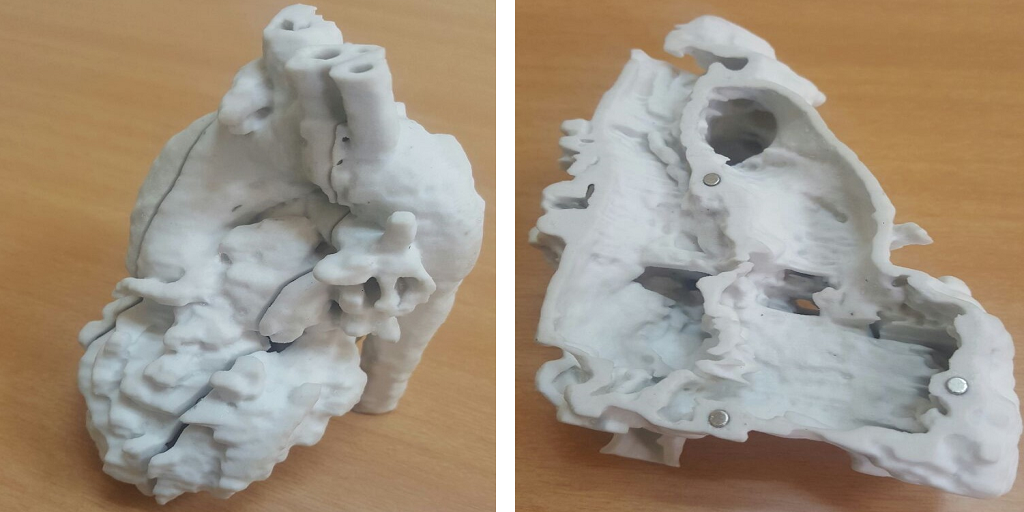
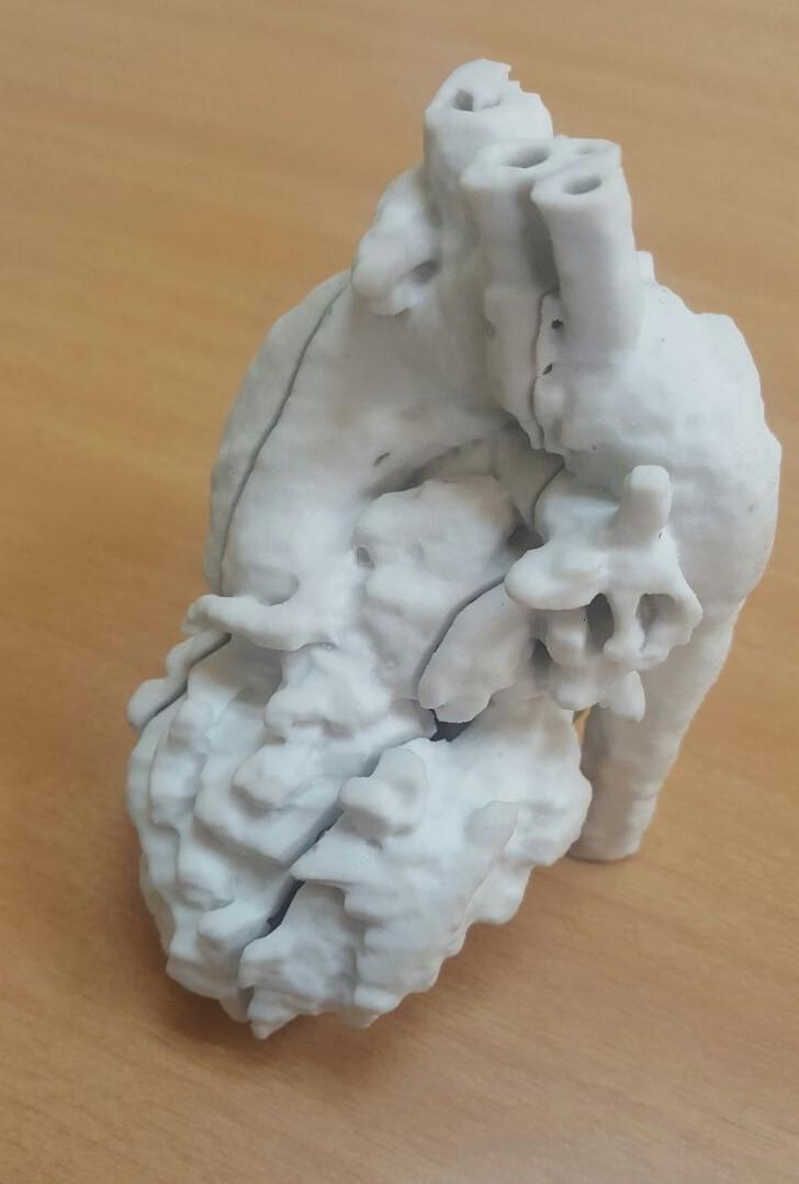
3D model of Sunita’s heart
When I started writing about 3D printing, I never expected that I would learn so much about medicine. I’ve learned a great deal about the heart, in particular – knowledge that’s both enlightening and frightening, as I never realized the incredible variety and complexity of conditions that can affect this critical organ. The bright side, though, is that the knowledge of these conditions comes with the knowledge that they can be fixed with the help of 3D printing, and that’s what blows my mind – the fact that surgeons can completely repair a heart so badly malformed as to be actually twisted, or a heart with its veins out of place. It wasn’t long ago that surgeries like these were considered too risky to even be attempted, but with 3D printed surgical models that allow surgeons to study and plan before operating, these procedures can not only be performed, but can turn even the most damaged hearts into healthy ones.
Today I learned about yet another heart condition that sounds too complicated to be imagined, much less survived. Sunita (name changed), the five-year-old daughter of a fisherman from Palghar, India, suffered from Ventricular Septal Defect (VSD), meaning that a large hole sat between the two lower chambers of her heart. Her condition was further complicated by the fact that her pulmonary artery was too narrow and abnormally positioned, leading to a diagnosis of “double outlet right ventricle with malposed great arteries with a large inlet Ventricular Septal Defect and severe Pulmonary Stenosis.”
Sunita’s parents were understandably frightened by her condition, but they were even more frightened by the risks that a surgical procedure would present. When she turned five, however, she began exhibiting more severe symptoms such as shortness of breath, sweating and pallor, and treatment could no longer be put off. On the suggestion of their family doctor, Sunita’s parents brought her to Fortis Hospital, where they were referred to Dr. Vijay Agarwal.
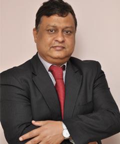
Dr. Vijay Agarwal
“Sunita’s condition was rare due to the complications involved. Her Aorta was at wrong place – instead of being above the left ventricle, it was placed above the right ventricle,” Dr. Agarwal explained. “And this was further complicated by her narrowed Pulmonary Arteries. When we first assessed the patient in our OPD, we realized that we could approach Sunita’s case in two totally different ways. In the first approach, we could have performed two separate surgeries to fix two different conditions. Though this plan had low risk, the heart defects would not be corrected fully with this approach. The second option would be corrective, but radical, in which the heart would be opened up and the base of the Aorta would be cut off from the heart. The Aorta would then be re-stitched over the correct ventricle (left ventricle). The remaining steps would be routine. This surgery is called ‘Nikaidoh Procedure.’”
The Nikaidoh Procedure, due to its risk and complexity, is very rarely performed, but the surgeons at Fortis Hospital decided to go ahead with it after evaluating the pros and cons of both options. Luckily, Dr. Agarwal has a history of successfully performing risky pediatric heart surgeries – thanks in no small part to the use of 3D printed surgical models. As in the case of other pediatric patients they had operated on, the Fortis surgical team took CT scans of Sunita’s heart and used them to create a 3D model, which was then printed.
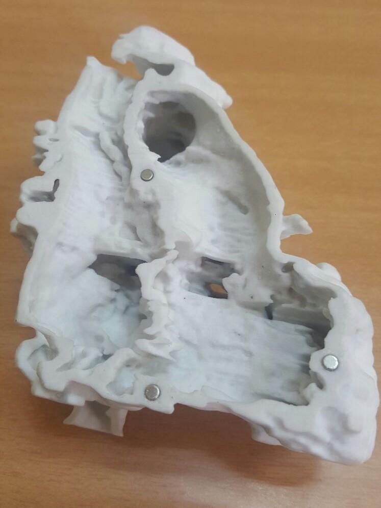
“With a precise 3-D model of the heart, we were certain about the problem areas and were able to plan out the surgery with greater confidence of success,” said Dr. Swati Garekar, a pediatric cardiologist at Fortis Hospital. “Given its complexity and risks involved, the Nikaidoh Procedure is rarely used in the city. But with our experience of handling similar cases in the past, the state-of-the-art facilities at Mulund’s Fortis Hospital and with the help of 3D Print model of the heart, we were able to manage risks and performed the surgery successfully.”
The surgery was performed in January, and Sunita is recovering well. Her life expectations – both length and quality – are now the same as those for any other child. These are the stories I’ll never get tired of hearing. What are your thoughts on the impacts technology is making in medicine and surgery? Discuss in the 3D Printed Heart Model for Sunita forum over at 3DPB.com.