If you go back just 2 years, it was almost unheard of to see media coverage of 3D printing technology coming to the aid of doctors in the operating room. Now, it seems as though every week there is a new “breakthrough” surgery that takes place with the help of 3D printing. Most of the stories that are reported, seem to originate either from the US, Europe or China. These are the places with the most prominent media coverage, and areas on the globe that 3D printing has really begun to gain a strong foothold in. However, one man, named Jorge Vicente Lopes da Silva, has been helping doctors and surgeons in Brazil use 3D printing and scanning technologies for years, yet you don’t see these stories in the mainstream media.

A scan taken of a 12-year-old’s lower body, prior to creating a 3D printed model.
Silva, the chief of the Tridimensional Technologies Division of the Center for Information Technology Renato Archer (CTI), has been aiding doctors and surgeons in the creation of 3D models for years now, even though the media just recently has begun to massively cover this type of medical technology.
“CTI is IT research center from the Science and Technology Ministry of Brazil, developing and integrating solutions for better surgical planning,” Silva told 3DPrint.com. “We have also many other applications like Egyptology, paleotology, etc. Also we developed an experimental technology for about 3500 cases with more than 100 hospitals in Brazil and some from Latin America.”
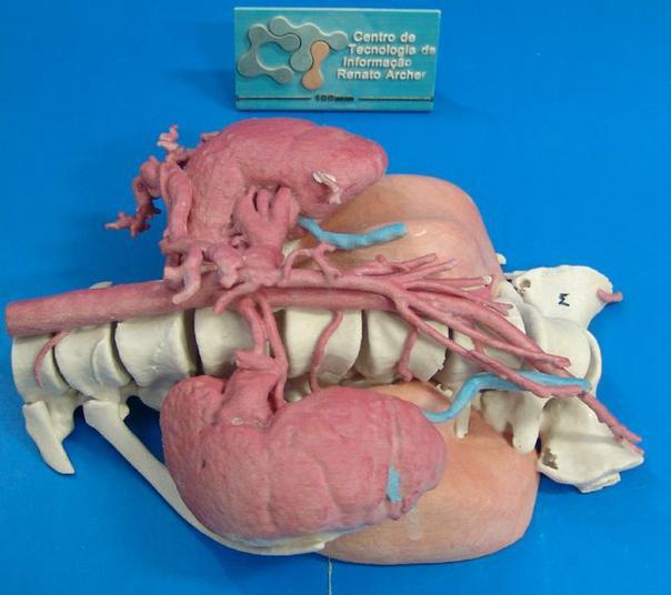
3D printed model of tumor, kidneys, arteries & more.
One particularly interesting case, which Silva is quite proud of, is a surgery that was performed at the Centro Médico, Campinas, in São Paulo, Brazil.
A 12-year-old girl had developed a rather ominous tumor, which was very large in size and located in a position where surgery carried many risks. Each and every year, many tumors fall under the unfortunate category of being consider “inoperable”. This is because they are surrounded by healthy tissue, organs, veins, and arteries, that if damaged in the removal process, could be life-threatening. In this little girl’s case, there was another risk involved, and that was the tumor’s proximity to her spine.
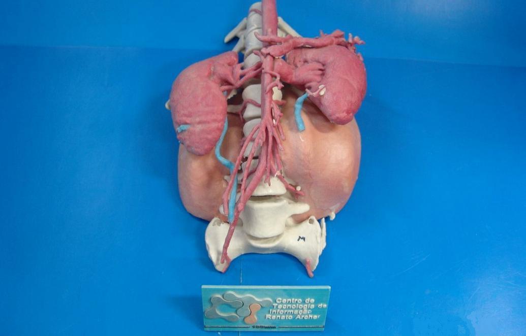
3D printed model that aided surgeons in 12-year-old girl’s tumor and vertebrae removal.
It was determined that the girl would need to have the tumor, along with 5 of her vertebrae removed. The tumor was located in a position very close to vital arteries and her kidneys. This surgery was an extremely risky one, one that most surgeons would probably consider high risk. Dr. Jose Carlos Barbi Gonçalves, however, had CTI at his side. With their help, Gonçalves used 3D technologies to carefully plan the surgery. He was assisted by both virtual and 3D printed models of the girls spinal column, tumor, arteries and kidneys. It allowed him to plan the “perfect” surgery to remove the tumor.
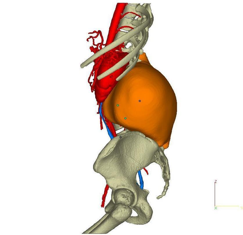
Virtual 3D model of the tumor.
The surgery ended up being very successful, in a large part due to the 3D printed models that Gonçalves was provided with. The models allowed him to see a 3-dimensional representation of the entire situation, so that he could carefully maneuver around vital body tissue, and significantly reduce the risk of damaging them.
This is just one of many ways CTI has come to the aid of surgeons performing surgery in South America. Let us know what you think of this incredible technology in the 3D Printed surgery forum thread on 3DPB.com.