Any number of sources will tell you that when dealing with cancer, early detection is a key element to increasing the chances of survival. In a paper published in the most recent issue of Transactions the journal of the Hong Kong Institution of Engineers (HKIE), a research team from the Chinese University of Hong Kong’s Institute of Precision Engineering described a device they have developed to aid in the early detection of cancers in the gastrointestinal tract.
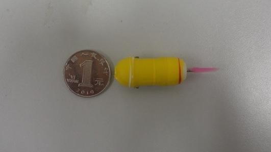
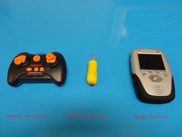
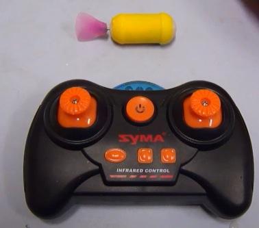
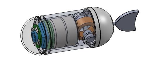
The miniature machine, composed of electronics and a wireless video camera enclosed in a 3D printed shell, is named the Tadpole Endoscope because the mechanics of its movement are based on those used by a tadpole to propel itself through its watery environment. The size of a large pill, the tadpole is introduced into the patient’s gastrointestinal tract by swallowing. Once inside, it can be directed via remote control and send back to a screen images of its surroundings.
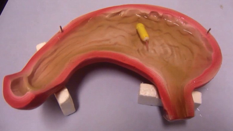
To date, the tadpole has not been released into the GI tract of any human beings. Instead, it has been undergoing rigorous testing, first in a model of the stomach and then later in the stomach of a pig. The successful test of the model with the propulsion system takes it one step closer to being ready for human use, but there are still a great deal more tests to undergo before then. What are your thoughts on this possible breakthrough within the cancer diagnostic space? Discuss in the 3D Printed Tadpole Endoscope forum thread on 3DPB.com.