
![]()
But heart specialists at Spectrum Health, a group of 12 not-for-profit hospitals in West Michigan and considered one of the best healthcare providers in the country, has just 3D printed the first model of a patient’s heart using combined data from imaging techniques. The hybrid 3D printed model has significantly more detail than models created using standard techniques. Spectrum Health is the first healthcare provider to develop a process to combine virtual data from multiple imaging scans.
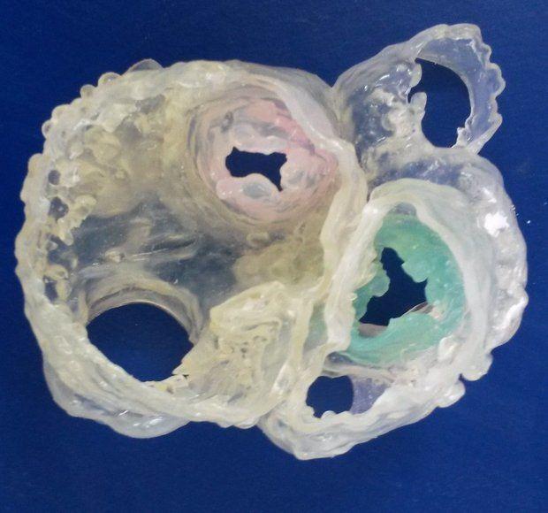
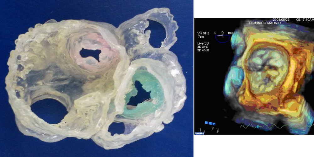
3D heart model created by combining CT scan data and 3D transesophageal echocardiography data
Typically when a surgeon needs a 3D model of a patient’s heart they use one of several methods available to them. In the last year we have seen hearts 3D printed using data from CT scans, MRI data and recently even an ultrasound process called 3D echocardiography. This 3D model printed by Spectrum Health combined the data gathered from a CT scan and using 3D echocardiography. Spectrum Health is also further developing the new process to incorporate highly detailed data from an MRI scan.
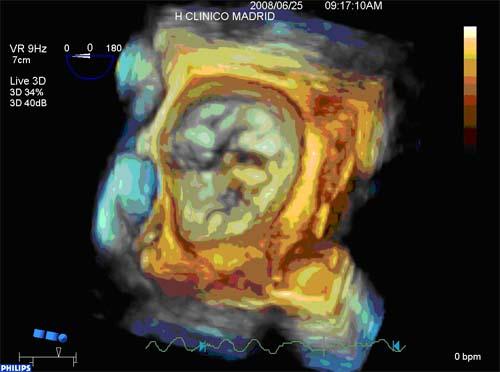
Mitral valve prolapse scan collected using 3DTEE imaging
“Hybrid 3D printing integrates the best aspects of two or more imaging modalities, which can potentially enhance diagnosis, as well as interventional and surgical planning. Previous methods of 3D printing utilize only one imaging modality, which may not be as accurate as merging two or more datasets,” cardiac sonographer and the lead author of the proof-of-concept study Jordan Gosnell explained to Mlive.com.
Each of the various imaging techniques used in this hybrid process has different strengths, and by combining data from multiple techniques doctors can create an extremely detailed 3D model of the patient’s heart. The CT scan is used to collect highly detailed data of the exterior anatomy of the heart. The inside of the heart is visualised using an MRI, which can also generate detailed scans of its musculature. And finally the 3D transesophageal echocardiography (3DTEE) process focuses specifically on the heart valves, the primary location of the type of defects that the process was developed to correct.

Dr. Joseph Vettukattil, interventional cardiologist with Spectrum Health’s Congenital Heart Center
The final proof-of-concept study for this process is being presented this week at CSI 2015 (The Catheter Interventions in Congenital, Structural and Valvular Heart Disease Convention) in Frankfurt, Germany. The presentation will be handled by the study’s senior author, Dr. Joseph Vettukattil, one of the leading heart specialists in the world. He has also helped develop the process of generating 3D models using 3DTEE, specifically for use with patients diagnosed with congenital heart disease. While the new process has already proven to be effective, Dr. Vettukattil does say that they still need to conduct additional research before determining the role of these hybrid 3D models in making surgical decisions. You can discuss this new medical advancement over in our 3D Printed Heart Model Using Multiple Imaging Techniques forum thread at 3DPB.com.