
In the past couple of years, we have finally begun to see 3D technology used in various hospitals around the world. 3D technology combines the advances of 3-dimensional imaging with that of 3D printing, to create tangible replicas of portions of a person’s body. China is one country that has been in the news a whole lot lately for their ever-increasing reliance on 3D technology, and this is seen yet again in today’s incredible story.
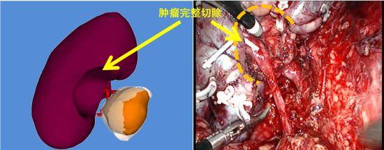
Earlier this month, doctors at Xiangya Hospital of Urology, Central South University in China, were able to not only successfully remove a tumor from a 60-year-old woman’s kidney, but in doing so they were also able to save that kidney — a feat that wouldn’t have been possible without the help of 3D printing.
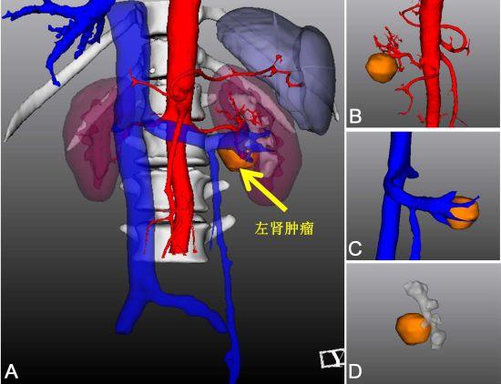
60-year-old Lee was diagnosed with a left renal tumor. It was located right next to the renal hilum which consists of many vital arteries and veins. Typically a surgery to remove this type of tumor would not be possible. In most cases the entire kidney must be removed, in a procedure referred to as a “radical nephrectomy”. This is done in order to reduce the great risks of potentially cutting an important artery or vein in the process, or of having the patient suffer from IRI (ischemia-reperfusion injury).
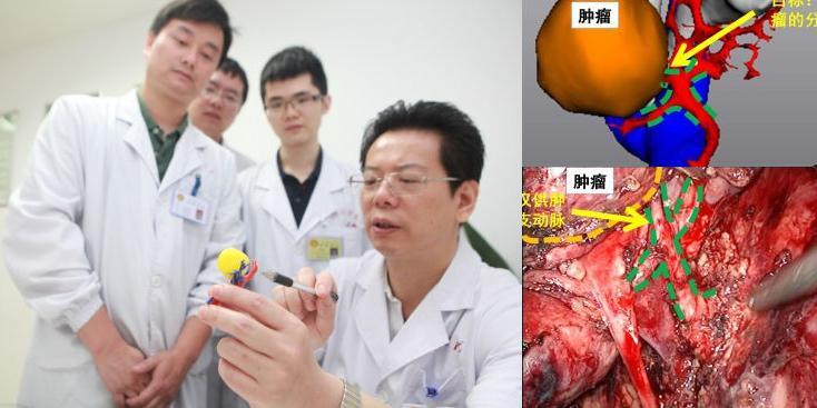
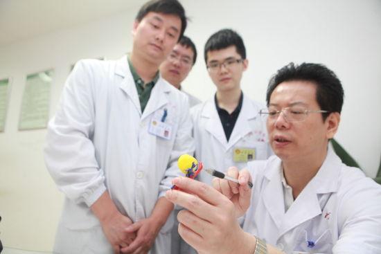
Xiangya Hospital of Urology Physician, Qi Lin, decided to use some of today’s most advance 3D technology to assist in surgical planning — technology that would allow the tumor to be removed while also allowing Lee to keep her kidney. In doing so, doctors took accurate 3-dimensional CT scans of Lee’s kidney, the tumor and the surrounding tissue, before creating a series of 3D printed replicas. Using these replicas, doctors were able to easily distinguish exactly where Lee’s renal artery, as well as the smaller branches of that artery was located, and ultimately come up with a precise plan for surgery.
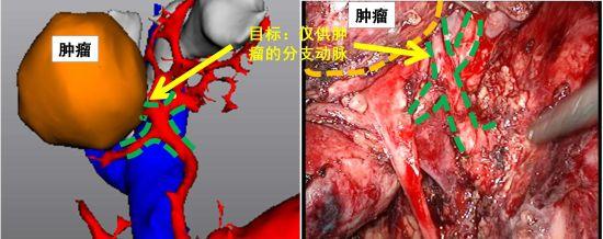
“With this new 3D technology, when aided by the surgeon, it creates a situation where ‘seeing is believing’. This is true from multiple dimensions, and allowed us to pinpoint the tumor, the arteries and the surrounding kidney tissue before surgery, and then decide on the correct path and operation to perform,” Professor Qi Lin explained.
All in all, the surgery was deemed a success. Lee is expected to make a full recovery, and Qi Lin’s team will certainly consider the use of 3D technology in future surgeries as well.
What do you think about this unique means of removing a tumor from a very dangerous spot in the human body? Discuss in the 3D printed kidney tumor model forum thread on 3DPB.com.