With less than 24 hours to go before he faced heart surgery to relocate heart vessels which were constricting and compressing his wind pipe and esophagus, tiny not-quite-two-year-old Liam Summers waited as Washington University cardiologist Dr. Peter Manning and his surgical team examined a precise 3D replica of Liam’s heart.
Liam’s father, Mike Summer, said seeing the 3D printed model of his son’s heart helped him understand the procedure his boy was facing at the St. Louis Children’s Hospital in Missouri.

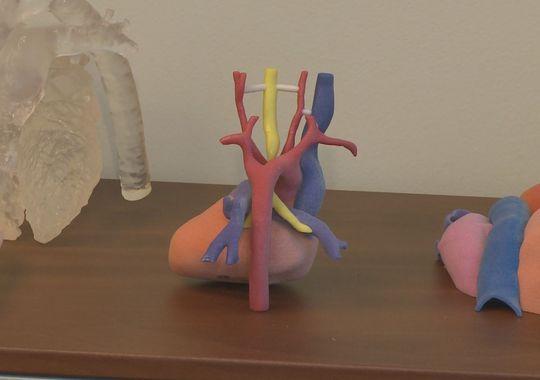
It’s all in a day’s work for cardiologists like Dr. Manning as 3D printed models are becoming commonplace tools in the process of medical care. They make it simpler for patients and families to grasp the details of complex medical procedures, and they provide medical practitioners with invaluable opportunities to practice and prepare for their work in the operating room.

Dr. Peter Manning
The 3D printing technology in play in Liam’s case resulted from combining MRI images and a 3D printer to create a life-sized replica of the boy’s heart and the network of vessels that were the cause of the problems he had breathing and swallowing.
Dr. Shafkat Anwar, a member of the cardiology team who handled Liam’s case, worked with 3D Systems to develop the model heart.
“It is something else for us to be able to put out the exact replica of the patient’s anatomy and hand that to someone who’s going to be doing the operation, and say ‘this is what you’re going to be facing in the operating room,'” Dr. Anwar told KSDK.
As he held a model of Liam’s heart, Dr. Manning explained how the model impacted the surgical team and the resulting procedure.
“This helps us recognize why the child might have breathing problems,” Manning said. “There’s an area that looks like narrowing of the trachea. We know the esophagus goes right through there, so every time that he swallows things are likely to get hung up there as well,” said Dr. Manning.
That Thursday morning marked the occasion of 20-month-old Liam’s fifth surgery. Only a scant few minutes after the two-and-a-half hour surgery was complete, Dr. Manning said the 3D printed of model of Liam’s heart proved critical to the success of the operation.
“The 3D model and the imaging we could create with the MRI gave us a very accurate picture that let us get the dissection done more quickly and less trauma to tissue in there,” Manning said. “He went to sleep fine and everything went real smoothly. We got in there and the anatomy was just like we expected. Swallowing stuff, I would guess, should be noticeably better within the next couple of weeks.”
Liam’s surgery was yet another example of how doctor’s are using 3D printed models to inform themselves and their teams of what to expect during delicate surgeries.
What do you think of how doctors use 3D printing to help patients with a wide range of medical issues? Let us know in the 3D Printed Model of Two-Year-Old’s Heart forum thread on 3DPB.com.